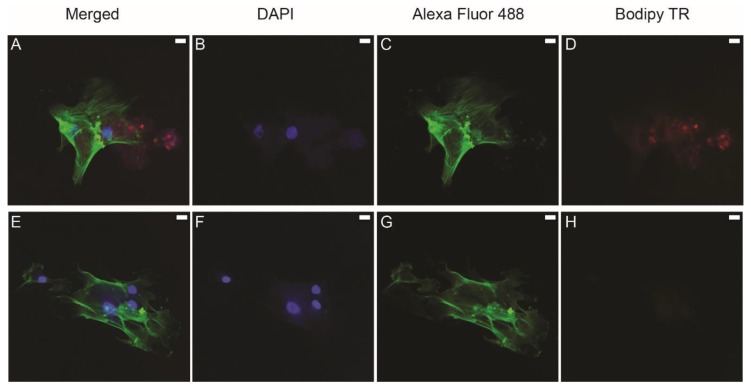
Figure 2.
Confocal immunofluorescent analysis of exosome uptake using DAPI to stain the nucleus (B,F), Alexa Fluor 488 to stain cytoskeletal actin (C,G), and BODIPY TR to stain exosomes (D,H). (A,E) are merged images. Labeled exosomes were isolated from the culture supernatants of dental pulp cells (DPCs). Human bone marrow-derived mesenchymal stem cells (HBMMSCs) were cultured with labeled exosomes. Red dots (A,D) represent exosomes. The red signal was not expressed in the negative control where non-labeled exosomes were added to the cells (E–H). Scale bar represents 20 µm.

