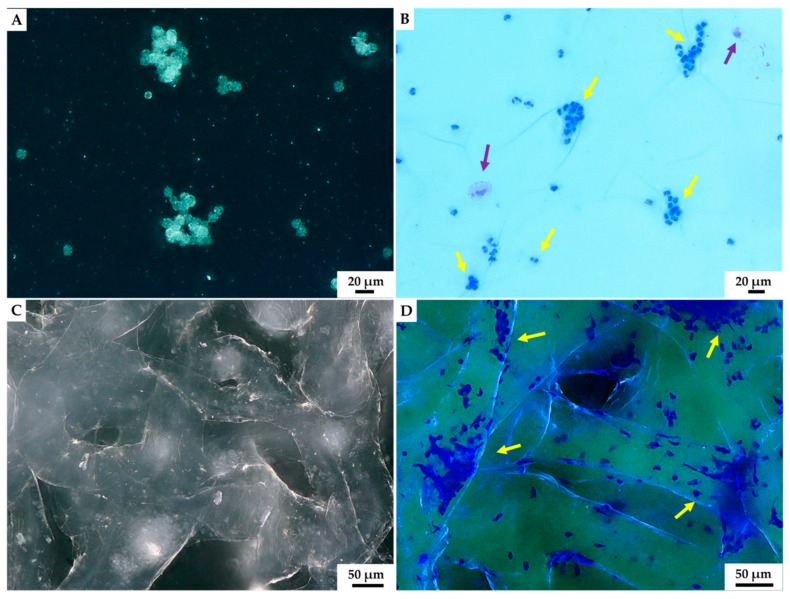
Figure 7.
Light microscopy imagery. Characteristic aggregates of hemocytes present in isolated hemolymph of C. aspersum snail was observed both without (A) and using eosin and methylene blue staining (B) on the glass slide. The formation of hemocytes-based clumps before (C) and after staining by eosin and methylene blue (D) became visible on the surface of A. archeri chitinous scaffold 24 h after the immersion in hemolymph. Two hemocytes types could be distinguished after staining: most dominant granulocytes (yellow arrow) and single hyalinocytes (violet arrow).

