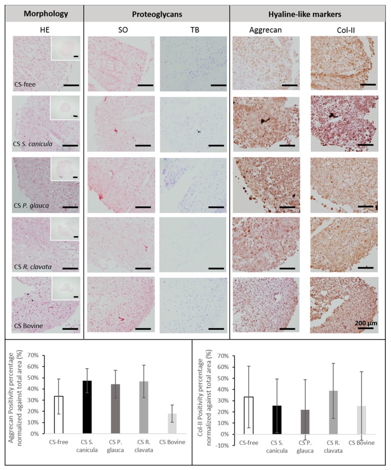
Figure 3.
Histological analysis for morphology (haematoxylin and eosin, HE), proteoglycans (toluidine blue, TB) and sulfated glycosaminoglycans (GAGs) synthesis (safranin-O, SO), and immunolocalization of collagen-type II (Col-II) and aggrecan (scale bar = 200 µm), was performed in osteoarthritic bone marrow mesenchymal stromal cells (BM-MSCs) pellets, after 14 days, in chondrogenic medium supplemented with 100 µg/mL CS extracted from fish (Prionace glauca, Raja clavata and Scyliorhinus canicula) and bovine sources or CS-free. Immunopositive aggrecan (bottom, left) and collagen type-II (bottom, right) percentage area were normalized against cell-pellets total area (n = 3).

