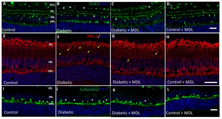
Figure 6.
Treatment with MDL 72527 reduced neurodegeneration in the diabetic retina. (A–D) Immunostaining of retinal cryostat sections using the ChAT antibody showing loss of amacrine cells in the diabetic retina (16 weeks), compared to control. (E–H) Immunostaining using PKCα, a marker for rod bipolar cells demonstrates the presence of degenerating axons and synaptic ends in the diabetic retina. (I–L) Immunofluorescence images showing the loss of horizontal cells in the diabetic retina by calbindin immunoreactivity. MDL 72527 treatment markedly reduced the neurodegenerative changes. Asterisks indicate areas of cell loss, and arrows indicate areas of axonal degeneration. N = 5–6 per group and representative images are presented. Scale bar 50 µm.

