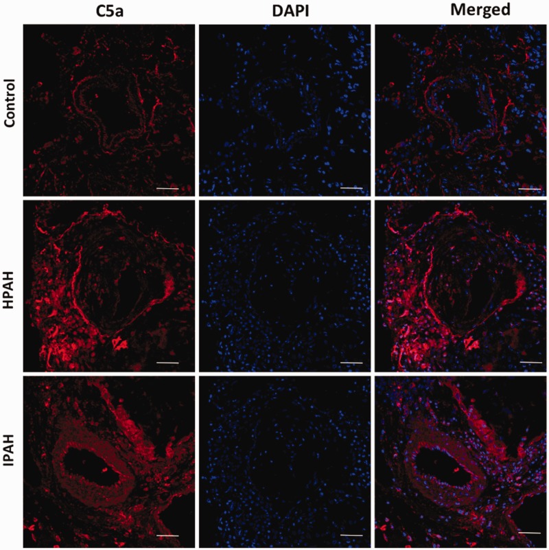
Fig. 6.
In PAH, C5a is increased in cells within pulmonary vessels and inflammatory cells surrounding these vessels: immunofluorescence staining for C5a (red color) is seen in inflammatory cells surrounding the pulmonary vessels and in cells of pulmonary vessels. Nucleus is stained blue (magnification 400×, scale bar 50 uM).

