Figure 4.

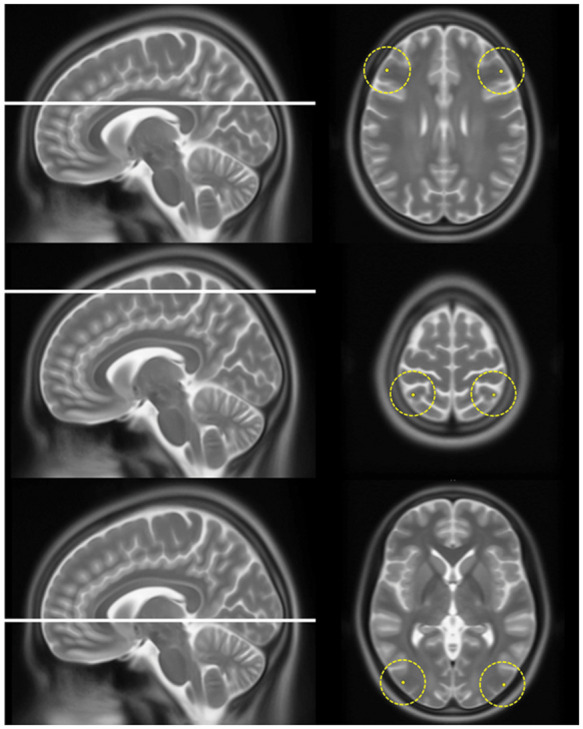
The six locations in the frontal, parietal and occipital lobe on which jPVS were graded. This figure shows the six locations (with a radius of 2 cm) on which jPVS were graded in all patients. The average score was used in the analysis.

The six locations in the frontal, parietal and occipital lobe on which jPVS were graded. This figure shows the six locations (with a radius of 2 cm) on which jPVS were graded in all patients. The average score was used in the analysis.