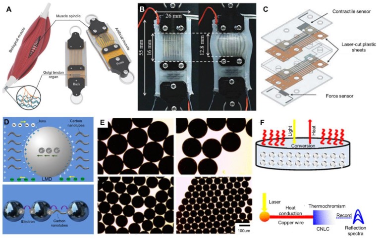
Figure 11.
Artificial muscles and liquid metal microsphere sensors. (A) Schematic comparison between biological muscle and artificial muscle, reproduced with permission from [136]. (B) The sensorized, flat, pneumatic artificial muscle at rest and inflated to 82.8 kPa, reproduced with permission from [137]. (C) Illustration of the contraction and pressure sensors, reproduced with permission from [137]. (D) The electrical environment of a liquid-metal microsphere in NaOH solution (above) and two methods of communicating for liquid-metal microspheres in a circuit (below), reproduced with permission from [77]. (E) Microscopy images showing liquid-metal microspheres produced by capillary-based microfluidics, reproduced with permission from [77]. (F) Schematic diagram of the photothermal conversion of liquid-metal microspheres and the near-infrared (NIR) sensor based on the liquid metal microsphere, reproduced with permission from [138].

