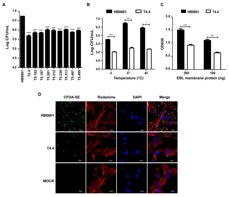
Figure 1.
M. bovis mutants with altered binding properties. (A) Binding of M. bovis mutants to EBL cells. For each mutant, the binding to EBL cells was determined by counting the number of CFU associated with cell monolayers following incubation with 108 CFU. Data are mean mycoplasma titers from three independent assays. Standard deviations are indicated. (B–D) Adhesion of HB0801 and Mbov_0503 knock-out mutant T4.4 to EBL cells. (B) Influence of the temperatures on the binding capacity. For each mycoplasma strain, the binding to EBL cells was determined by counting the number of CFU/mL associated with cell monolayers following incubation with 108 CFU. Data are mean mycoplasma titers from three independent assays. Standard deviations are indicated. (C) Quantification of M. bovis binding to EBL membrane proteins. Microplates were coated with 100 ng or 200 ng EBL membrane extracts; 107 CFU of each strain were used. (D) Visualization of CFDA-SE labeled mycoplasmas bound to EBL cells by laser scanning confocal microscopy. Actin filaments and nuclei were labeled with Rhodamine phalloidin and DAPI, respectively. PBS was used as a negative control (MOCK).

