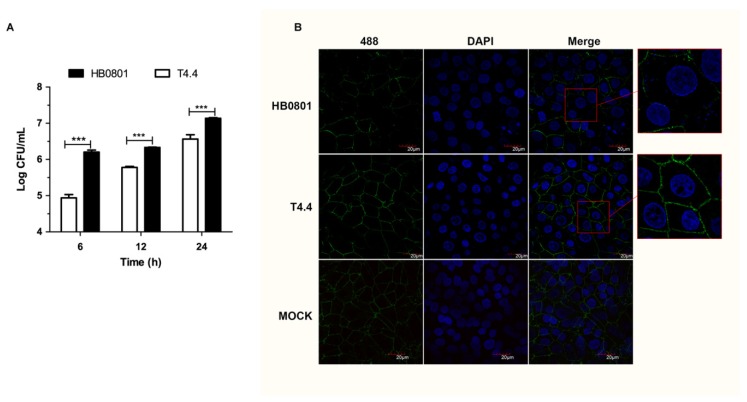
Figure 5.
Translocation of Mbov_0503 knock-out mutant across MDBK epithelial cell monolayers. (A) MDBK cell monolayers grown on Transwell chambers were infected with Mbov_0503 knock-out mutant (T4.4) or the parental strain (HB0801) at the apical side, and mycoplasmas that translocated to the basal chamber were enumerated by counting CFU. (B) Structural integrity of the tight junctions in MDBK cell monolayers infected with the Mbov_0503 knock-out mutant (T4.4) or the parental strain (HB0801). The tight junctions were visualized by laser scanning confocal microscopy using anti-ZO-1 and Alexia 488-conjugated anti-IgG antibodies (Green). The nuclei were labeled with DAPI (Blue). MOCK-infected MDBK cell monolayers were used as a negative control (MOCK). Images in red boxes were magnified for 2.5 times. P values are indicated by asterisks (*** p < 0.001).

