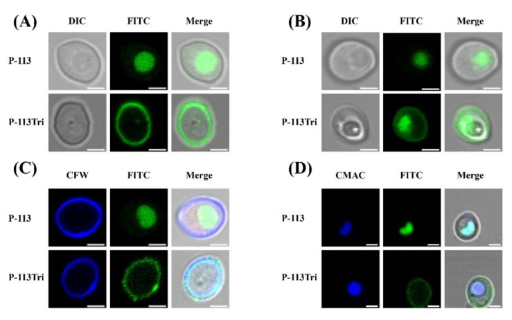
Figure 1.
Examination of the interactions of P-113 and P-113Tri with Candida albicans cells using confocal microscopy. (A) Differential interference contrast (DIC) and fluorescence images show that FITC-P-113 quickly gains entry into the cells within 5 min. (B) FITC-P-113 and a part of FITC-P-113Tri accumulated in vacuoles after 1 h of treatment. (C) FITC-P-113Tri was found to bind to the cell wall, as demonstrated by colocalization with calcofluor white. (D) The peptides inside the cells accumulated in vacuoles as demonstrated by colocalization with CMAC. Scale bar, 2 μm.

