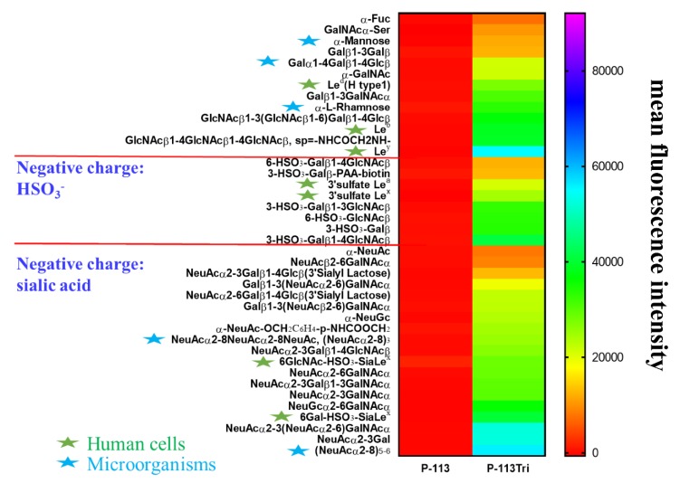
Figure 5.
Screening of carbohydrate targets for the peptides using a solution carbohydrate array. The array contained different terminal sequences of N-glycans, O-glycans, and glycosphingolipids of microbial cells and mammalian tissues. Briefly, donor beads (500 ng/well) and biotin-PAA-sugars were mixed with FITC-P-113 and FITC-P-113Tri. Subsequently, a mixture of acceptor beads, mouse anti-FITC antibody, and rabbit anti-mouse IgG antibody was added. The binding signals were analyzed with a PerkinElmer Envision instrument using AlphaScreenTM. The top 40 glycans with peptide binding are shown. Led (H type 1): Fucα1-2Galβ1-3GlcNAcβ, Leb: Fucα1-2Galβ1-3(Fucα1-4)GlcNAcβ, Ley: Fucα1-2Galβ1-4(Fucα1-3)GlcNAcβ, 3’sulfate Lea: 3-HSO3-Galβ1-3(Fucα1-4)GlcNAcβ, 3’sulfate Lex: 3-HSO3-Galβ1-4(Fucα1-3)GlcNAcβ, 6GlcNAc-HSO3-Sia Lex: Neu5Acα2-3Galβ1-4(Fucα1-3)(6-HSO3)GlcNAcβ, 6Gal-HSO3-Sia Lex: Neu5Acα2-3(6-HSO3)Galβ1-4(Fucα1-3)GlcNAcβ. Gal: galactose; GalNAc: N-acetylgalactosamine; Glc: glucose; GlcNAc: N-acetylglucosamine; NeuAc: N-acetylneuraminicacid; NeuGc: N-glycolylneuraminicacid. Green stars: glycans presented in human cells; Blue stars: glycans presented in microbial cells.

