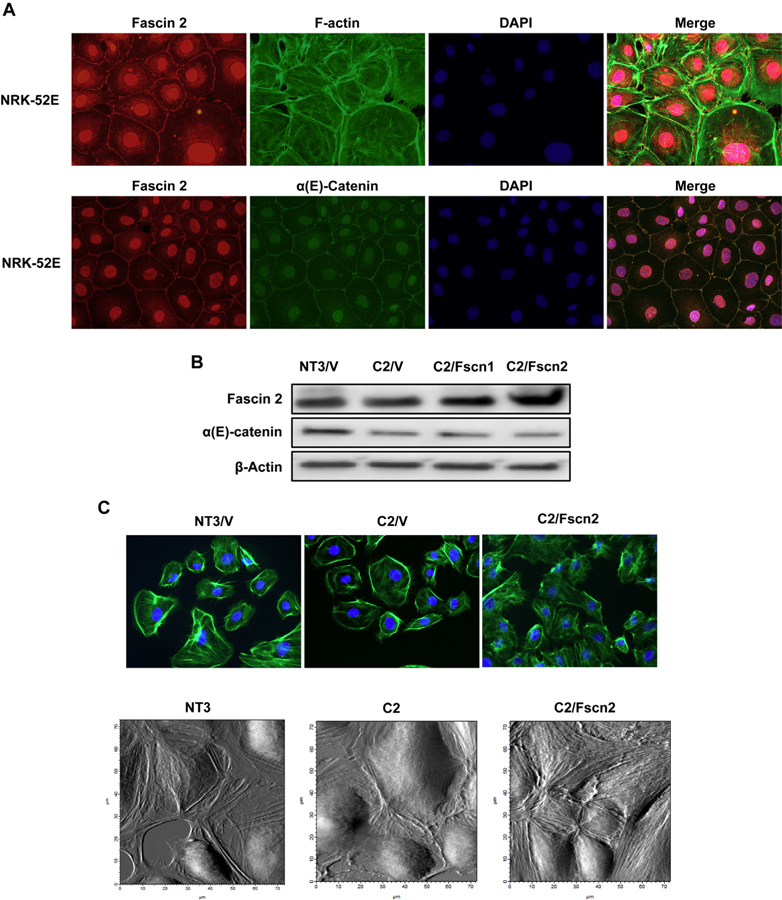
Fig. 2.
Fascin2 Colocalizes with α-Catenin and the Actin Cytoskeleton; Overexpression of Fascin2 Rescues Stress Fibers in C2Cells. A. Immunofluorescence images (60x) of NRK-52E cells treated with rabbit anti-fascin2 (red), FITC-phalloidin (green) or anti-α-catenin (green) and DAPI (blue) demonstrates that fascin2 colocalizes with both actin and α-catenin. B. Overexpression of fascin1 (C2/Fscn1) or fascin2 (C2/Fscn2) in C2 cells. C. Phalloidin staining demonstrates increased actin stress fibers in C2/Fscn2 cells, while the bottom panel shows stress fibers using cell surface scanning with AFM. (For interpretation of the references to colour in this figure legend, the reader is referred to the web version of this article.)

