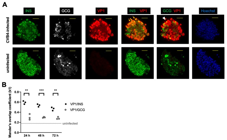
Figure 1.
Immunostaining of stem cell-derived β (SC-β) cells revealed abundant insulin (INS)- and glucagon (GCG)-positive cells with viral protein (VP1) predominantly colocalizing with INS. (A) The colocalization of green and red is shown as yellow. Arrows show examples of colocalization between VP1 and INS, and the arrowhead shows colocalization between VP1 and GCG. A representative cell cluster from each condition at 48 h post-infection with coxsackie B virus (CVB)4 (or control uninfected) is shown. INS = green, GCG = white or green, VP1 = red, Hoechst (DNA stain) = blue. Scale bar = 50 μm. (B) Mander’s overlap coefficients (k1) for VP1 and INS and for VP1 and GCG in infected cells are shown at the indicated time points following infection. VP1 and INS colocalized more frequently than VP1 and GCG. (** p < 0.01; *** p < 0.001, multiple t-test). The coefficient was significantly lower for uninfected controls for which the median value is shown as the horizontal dashed line (p < 0.0001, Mann–Whitney test). Each point represents one field of view.

