ABSTRACT
Background
Sepsis-induced myocardial dysfunction has implications on outcome. For lack of echocardiography in resource-limited settings, myocardial biomarkers may be an alternative monitoring tool.
Objective
This study was planned to explore the longitudinal behavior of creatine phosphokinase-MB (CPK-MB) in children with sepsis with and without shock, and its correlation with clinical and echocardiographic parameters over the first 10 days.
Design
Prospective observational study.
Setting
Tertiary care hospital in a lower-middle-income economy of South Asia.
Patients
Children (3 months to 12 years) with nonshock sepsis (NSS) (n = 40) and septic shock survivors (SSSs) (n = 40) after optimal resuscitation. Patients with catecholamine refractory shock, preexisting heart disease, and cardiorespiratory event within the past 1 month were excluded from the study.
Measurements and main results
Pediatric logistic organ dysfunction (PeLOD) score, vasoactive inotrope score (VIS), CPK-MB, and echocardiographic measures of myocardial function were recorded on days 1, 3, 7, and 10. Echocardiography was repeated at 1 month. Both groups were similar at baseline. The SSSs had higher CPK-MB (180 vs 53 IU/L; p < 0.001) and PeLOD score (2 ± 0.4 vs 11.7 ± 5.1, p < 0.001) on day 1 compared to the NSS children. More than half of the SSS and none of the NSS patients had myocardial dysfunction. Reduction in CPK-MB over 10 days correlated well with improvement in PeLOD (p < 0.01), VIS (p = 0.04), and echocardiographic measures of myocardial dysfunction (p < 0.05) among SSSs. At 1 month follow-up, all had normal echocardiography.
Conclusion
The SSSs had markedly elevated CPK-MB, and its fall paralleled the improvement in clinical status and myocardial dysfunctions. The CPK-MB could be a potential monitoring tool for septic cardiomyopathy in resource-limited settings.
How to cite this article
Baranwal AK, Deepthi G, Rohit MK, Jayashree M, Angurana SK, Kumar-M P. Longitudinal Study of CPK-MB and Echocardiographic Measures of Myocardial Dysfunction in Pediatric Sepsis: Are Patients with Shock Different from Those without? Indian J Crit Care Med 2020;24(2):109–115.
Keywords: Cardiac biomarkers, Creatine kinase-MB, Echocardiography, Myocardial dysfunction, Sepsis, Septic shock
INTRODUCTION
Sepsis and septic shock are continuum of the same spectrum.1 It continues to remain the leading cause of death among under-fives, especially in low- and middle-income economies.2,3 Tertiary care teaching hospitals of our country report mortality of >40%.4–7 Although septic shock patients commonly have myocardial dysfunction8–15 and is associated with high mortality,12 it is reversible by 7–10 days among survivors.16–18 Myocardial dysfunction is conventionally assessed by echocardiography. Its limited availability at bedside in resource-limited facilities, especially in pediatric services, is a cause of concern. An affordable, sensitive, and specific biomarker of myocardial dysfunction, which helps to prognosticate and monitor the septic shock management is the need of the hour. Early recognition of myocardial dysfunction may help in the use of inodilators for quick and improved outcome.
Experimental models of myocardial injury have shown significant rise in creatine phosphokinase-MB (CPK-MB) and cardiac troponins (cTns).19,20 In clinical studies, elevations in cTnI and cTnT are shown to correlate with myocardial dysfunction, duration of hypotension, vasopressor requirement, and mortality.9,10,18,21–23 However, there is paucity of data on CPK-MB in pediatric septic shock, especially with regard to its utility with serial monitoring and its correlation with echocardiographic findings. We hypothesized that CPK-MB would be elevated in children with septic shock due to myocardial injury. This prospective observational study was conducted to explore the longitudinal behavior of CPK-MB in children with sepsis with and without shock, and its correlation with myocardial dysfunction as assessed by clinician-performed echocardiography over the first 10 days.
MATERIALS AND METHODS
Clinical Setting and Patients
Clinical setting of our hospital and patient population is already described elsewhere.7 Children in age-group of 3 months to 12 years were enrolled over a period of 12 months (April 2010 to March 2011) from pediatric emergency room (PER) and pediatric intensive care unit (PICU). Nonshock sepsis was defined by the presence of features of systemic inflammatory response syndrome (SIRS) in a child with clinically suspected infection.24,25 Septic shock was defined per the standard guidelines.24,26 Patients in the NSS group were enrolled as soon as possible and patients in the septic shock group were enrolled once they were resuscitated optimally and met the end points of septic shock resuscitation.25 Only the SSSs were included for the final analysis of the study. A convenience sample of 40 consecutive patients each was planned for enrollment in the NSS and the SSS groups. This was a nonfunded dissertation work, to be completed in a limited period, of the postgraduate pediatric training program. It was difficult to screen and enroll all the eligible patients consecutively, despite the best efforts of the trainee (G.D.) due to her rotations through various clinical units of the Department of Pediatrics. Thus, details of excluded patients among the eligible ones could not be provided.
Children with catecholamine-resistant shock for initial 24 hours, healthcare-associated infections, congenital or acquired heart disease, myocarditis, high-frequency ventilation, immunosuppressed state, neurodevelopment delay, neuropathy/myopathy, genetic syndromes, congenital malformations, raised intracranial pressure, postoperative patients, and any cardiorespiratory event within 1 month of admission and deferred consent were excluded from the study. Written informed consent was obtained from parents or legal guardians. The study protocol was approved by the Departmental Review Board, the Institute Ethics Committee, and the Institute Thesis Committee.
Data Collection and Clinical Care of Patients
Demographic and clinical data (including sepsis and septic shock criteria, PeLOD score, and VIS)27 of all enrolled patients were recorded. Patients were resuscitated and stabilized in PER the recommended guidelines25 followed by transfer to PICU as and when the beds became available. First VIS was calculated after initial resuscitation. The subsequent management was per the unit protocol. Patients were transitioned to pediatric ward care for ongoing management once their intensive care needs were taken care of.
All patients underwent transthoracic echocardiographic evaluation of the ventricular function within 24 hours of NSS or septic shock diagnosis (after having achieved hemodynamic stabilization with fluids and inotropes) and again on days 3, 7, 10, and 1 month following discharge. Echocardiographic examination was performed using the portable ECHO machine (ACUSON Cypress®, Cleveland, OH, USA) with 7 Hertz cardiac probe by the postgraduate pediatric trainee (G.D.) trained in pediatric cardiology unit for 1 month before the beginning of the study. She was trained in performing echocardiography specifically concentrating on the two-dimensional (2-D) echocardiography, M-mode, and spectral Doppler echocardiography techniques with specific emphasis on determining the left ventricular (LV) systolic and diastolic functions under a pediatric cardiologist (MKR). The person performing echocardiography was not involved in the clinical management of patients.
The LV systolic function was assessed by the LV ejection fraction (LVEF), fractional area change (FAC), and fractional shortening (FS). The LVEF (as per Simpson's rule) and FAC were measured in 2-D echocardiography by apical four-chamber view and FS by the standard M-mode. For assessing the LV diastolic dysfunction, E/A ratio (EAR) [ratio of peak velocity flow in early diastole (E) to the peak velocity flow in late diastole caused by atrial contraction (A)] was calculated in the apical four-chamber view by pulsed Doppler across the mitral valve. Each parameter was recorded thrice and their average value was taken as the final. All the recordings were saved in a computer. These were later reviewed and parameters were calculated by a pediatric cardiologist (M.K.R.). Lower limit of normal values for LVEF, FAC, and FS were considered to be 55%, 36%, and 25%, respectively.
Serum CPK-MB isoenzyme and C-reactive protein (CRP) were measured serially on days 1, 3, 7, and 10. The CPK-MB was measured by chemiluminescent enzyme-labeled immunometric assay method (DPC Immulite) and a value >25 IU/L was considered elevated. Serum cTnT card test [a one-step in vitro qualitative card test based on immunochromatography (sandwich immunoassay) technique] was done on day 1 only and not serially due to limitation of funds. Storage temperature of the card device was 2–8°C. A positive test indicated a serum level of >0.08 ng/dL. Trend and correlation between myocardial dysfunction (as assessed by LVEF, FAC, FS, and E/A ratio) and CPK-MB in NSS patients and SSSs during acute phase (days 1, 3, 7, and 10) and 1 month after discharge from hospital were the primary outcome measures, while the correlation of longitudinal trends of CPK-MB with CRP, PeLOD score, and VIS in the two groups formed the secondary outcomes.
STATISTICAL ANALYSIS
Data analysis was performed using “R” version 3.4.428 including additional packages like ggplot2,29 CAR,30 pROC31, and t-series.32 Chi-square (X2) test (or Fisher's exact test) and t test were used for comparing categorical variables and continuous variables, respectively. Longitudinal change in the biochemical and echocardiographic parameters were analyzed by repeated-measures analysis of variance, assuming departure from sphericity for all the comparisons and employing Greenhouse–Geisser correction. Augmented Dicky–Fuller (ADF) test for cointegration was used to analyze longitudinal correlation of CPK-MB with echocardiographic, clinical, and biochemical parameters despite knowing that our time points were not optimally dense. Furthermore, the ADF test requires data set with at least five time points. As 1-month CPK-MB values (i.e., the fifth time point data) were not available for SSSs, the 10th day CPK-MB values of NSS patients were imputed as 1-month CPK-MB values for SSSs with the assumption that SSSs are likely to have normal CPK-MB values by 1-month as was the case among NSS patients by 10th day. Receiver operating characteristic (ROC) curves were constructed to evaluate CPK-MB as the predictor of various echocardiographic measures, and the cutoffs for CPK-MB were calculated. DeLong method was used to compute area under curve (AUC) with 95% confidence interval (CI).
RESULTS
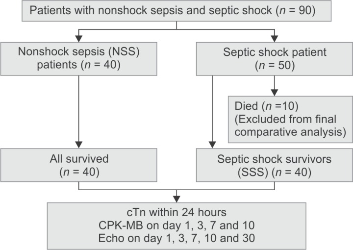
A total of 90 patients were enrolled during the study period. Forty patients fulfilled the criteria of clinically suspected infection, SIRS, and without hemodynamic compromise; these patients formed the NSS group. Fifty patients fulfilled the criteria of septic shock, of which 10 patients died mostly by 48–72 hours of admission. As the primary objective was to compare the trend of myocardial dysfunction in NSS patients with that of septic shock patients during acute phase and after 1 month follow-up, only survivors of septic shock were included for final analysis. The SSSs consisted of 40 patients. Data on nonsurvivors (n = 10) were provided for comparison (Flowchart 1).
Flowchart 1.
Study flow diagram
Demographic, nutritional, and clinical details are shown in Table 1. Septic shock patients were relatively younger than NSS patients. Pneumonia was the most common diagnosis followed by disseminated staphylococcal sepsis and central nervous system infections. Number of dysfunctional organs, PeLOD score at admission, and hospital stay were significantly higher in SSS compared to that in NSS patients. Mean ± standard deviation (SD) of maximum doses of various vasoactive drugs used among SSSs were as follows—dopamine (19.3 ± 2.9 μg/kg/minute), dobutamine (16.4 ± 4.5 μg/kg/minute), adrenaline (0.2 ± 0.7 μg/kg/minute), noradrenaline (0.2 ± 0.8 μg/kg/minute), and milrinone (0.8 ± 0.2 μg/kg/minute). Number of SSSs who were given various vasoactive inotropic drugs on day 1 was as follows—dopamine (n = 36), dobutamine (n = 22), adrenaline (n = 26), noradrenaline (n = 8), and milrinone (2). Compared to the SSSs, nonsurvivors of septic shock required significantly higher VIS (63.0 ± 16.4 vs 36.9 ± 19.4; p < 0.001) at admission. The later also had higher PeLOD score (15.3 ± 4.8 vs 11.7 ± 5.1; p = 0.059) at admission, though the difference could not reach statistical significance.
Table 1.
Demographic and clinical characteristics of patients
| Characteristics | Nonshock sepsis (NSS) (n = 40) | Septic shock (n = 50) | |
|---|---|---|---|
| Septic shock, survivors (SSSs) (n = 40) | Septic shock, nonsurvival (n = 10) | ||
| Age (months), median (IQR) | 43 (14.3–67.5) | 24 (8.8–62.5) | 18 (8–54) |
| Age | |||
| <1 year, n (%) | 8 (20%) | 12 (30%) | 4 (40%) |
| 1–5 years, n (%) | 15 (37.5%) | 16 (40%) | 3 (30%) |
| >5 years, n (%) | 17 (42.5%) | 12 (30%) | 3 (30%) |
| Sex ratio (M:F) | 31:9 | 23:17 | 7:3 |
| Severe acute malnutrition, n (%)a | 3 (8) | 4 (10) | 0 (0) |
| BMI, median (IQR) | 14.8 (14–16) | 14.7 (13.4–16.3) | 15 (14–16.8) |
| Primary diagnosis | |||
| Pneumonia, n (%) | 15 (37.5) | 26 (65) | 5 (50) |
| CNS infections, n (%) | 8 (20) | 5 (12.5) | 2 (20) |
| Disseminated staphylococcal | 3 (30) | ||
| Sepsis, n (%) | 7 (17.5) | 7 (17.5) | |
| Enteric fever, n (%) | 3 (7.5) | 0 (0) | |
| Others | 7 (17.5) | 2 (5) | |
| Total number of dysfunctional organs at admission, mean (SD)b | 0.8 ± 0.3 | 3.1 ± 0.9 | 3.4 ± 0.8 |
| PeLOD score at admission, mean (SD)b | 2 ± 0.4 | 11.7 ± 5.1 | 15.3 ± 4.8 |
| Patients who received mechanical ventilation, n (%) | 19 (47.5%) | 20 (50%) | 10 (100%) |
| Duration of mechanical ventilation in days, mean (SD) | 6.3 ± 3.1 | 6.6 ± 2.9 | Till death |
| Patients who received inotropes, n (%)b | 0 (0%) | 40 (100%) | 10 (100%) |
| VIS (mean ± SD) on day 1b,c | 0 | 36.9 ± 19.4 | 63.0 ± 16.4 |
| Duration of inotropes in days, mean (SD) | – | 6.4 ± 2.5 | Till death |
| Hospital stay (days) (mean ± SD)b | 10.8 ± 2.6 | 15.2 ± 4.3 | 2.7 ± 1.1 |
aNational Center for Health Statistics (NCHS) median weight was used as reference; bSignificant difference between NSS patients and SSSs, p < 0.001; cSignificant difference between septic shock survivors and septic shock nonsurvivors, p < 0.001
IQR, interquartile range; PEM, protein energy malnutrition; BMI, body mass index; PeLOD score, pediatric logistic organ dysfunction score; VIS, vasoactive inotrope score; SD, standard deviation, NA, information not available
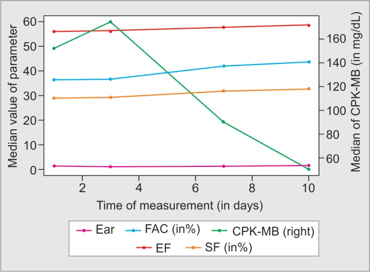
Table 2 provides details of biochemical and echocardiographic parameters at enrollment. The CPK-MB was elevated (>25 IU/L) in all SSSs compared to only two thirds of NSS patients (100% vs 77.5%, p = 0.01). Twenty-eight children in SSS group had CPK-MB >100 IU/L, while 11 had >200 IU/L. On the other hand, only two patients had CPK-MB >100 IU/L in NSS group and none more than 200 IU/L. The mean (±SD) CPK-MB was significantly higher among SSSs compared to NSS patients (180.8 ± 110.2 vs 53 ± 21 IU/L; p ≤ 0.001); the same trend was observed with CRP as well (p < 0.001). The cTnT card test was positive only in two septic shock patients and none in the NSS patients. Echocardiographic parameters were normal in all NSS patients and did not change over 10 days’ period (Fig. 1). While these were significantly abnormal in SSSs; LVEF (range, 35.1–58.5%), FAC (range, 21.2–47.5%), SF (range, 13.8–38.9%), and E/A ratio (range, 0.76–2.45) being lesser than lower limit of normal in 25%, 45%, 15%, and 12.5% patients, respectively. The mean and SD of various echocardiographic parameters in SSSs with myocardial dysfunction (i.e., below the corresponding cutoffs) were as follows—LVEF (50.05 ± 6.1), FAC (33.4 ± 4.3), SF (19.0 ± 5.6), and EAR (0.84 ± 0.07). Twenty (50%) and 5 patients (12.5%) had systolic and diastolic dysfunctions, respectively, while 3 patients had both systolic and diastolic dysfunctions. Overall, 22 SSSs (55%) had at least one echocardiographic parameter of myocardial dysfunction. Echocardiographic parameters improved significantly among SSSs; almost all patients had normal LVEF, FAC, SF, and E/A ratio by day 10 (Fig. 1). The reduction in CPK-MB among SSS correlated significantly with improvement in LVEF (ADF statistic, −4.43; p < 0.01), FAC (ADF statistic, −9.34; p < 0.01), SF (ADF statistic, −4.15; p = 0.018), and E/A ratio (ADF statistic, −3.78; p = 0.037) during the study period (Fig. 2). Survivors and nonsurvivors of septic shock were similar in terms of their CPK-MB levels (180.8 ± 110.2 vs 161.9 ± 40.9; p = 0.5) as well as echocardiographic parameters (Table 2).
Table 2.
Biochemical and echocardiographic parameters in nonshock sepsis and septic shock patients on day 1
| Parameters | Nonshock sepsis (NSS) (n = 40) | Septic shock (n = 50) | p values (NSS vs SSSs) | |
|---|---|---|---|---|
| Septic shock, survivors (SSSs) (n = 40) | Septic shock, nonsurvival (n = 10) | |||
| Creatine phosphokinase-MB (IU/L), mean ± SD | 53.3 ± 28.1 | 184.3 ± 109.5 | 161.9 ± 40.9 | p < 0.001 |
| Creatine phosphokinase-MB >25 IU/L, n (%) | 31 (77.5%) | 40 (100%) | 10 (100%) | p = 0.002 |
| C-Reactive protein (mg/L), mean ± SD | 49.6 ± 32.2 | 110.8 ± 85.5 | NA | p = 0.001 |
| Positive cardiac troponin T, n (%) | 0 | 2 (5%) | 0 | p = 0.152 |
| Left ventricular ejection fraction, mean ± SD | 58.8 ± 2.7 | 54.9 ± 4.2 | 56.2 ± 1.6 | p < 0.001 |
| Left ventricular ejection fraction <55%, n (%) | 0 | 10 (25%) | 3 (30%) | p < 0.001 |
| Fraction area change, mean ± SD | 45.7 ± 3.9 | 36.2 ± 4.4 | 39.4 ± 3.9 | p < 0.001 |
| Fraction area change <36%, n (%) | 0 | 18 (45%) | 2 (20%) | p < 0.001 |
| Fractional shortening, mean ± SD | 31.8 ± 2.7 | 29.4 ± 4.4 | 27.6 ± 3.7 | p = 0.005 |
| Fractional shortening <25%, n (%) | 0 | 6 (15%) | 4 (40%) | p = 0.011 |
| E/A ratio, mean ± SD | 1.44 ± 0.23 | 1.38 ± 0.34 | 1.46 ± 0.32 | p = 0.35 |
| E/A ratio <1, n (%) | 0 | 5 (12.5%) | 1 (10%) | p = 0.021 |
Fig 1.
Line diagrams comparing trends in creatine phosphokinase-MB, C-reactive protein, and echocardiographic parameters between nonshock sepsis (NSS) patients and septic shock survivors during the study period
Fig 2.
Line diagram comparing trends of creatine phosphokinase-MB with echocardiographic parameters (EAR, p = 0.037; LV ejection fraction, p < 0.01; fractional area change, p < 0.01; SF, p = 0.018) among septic shock survivors during the study period
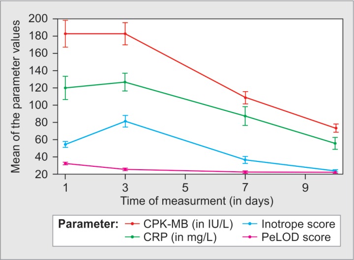
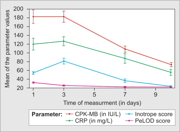
The CPK-MB, CRP, and PeLOD score had also gradually and significantly reduced over 10 days (p < 0.01) among SSSs, the most significant change being after day 3. The VIS first increased till day 3 (from 37 ± 14 on day 1 to 68 ± 49 on day 3) only to decrease significantly by day 7 (17 ± 29) (p < 0.01) (Fig. 3). By day 7, cointegration (ADF) test revealed a significant correlation between reduction in CPK-MB and reduction in PeLOD score (ADF statistic, −85.95; p < 0.01) and VIS (ADF statistic, −3.66; p = 0.04) over the study period. However, reduction in CPK-MB was not significantly correlated with reduction in CRP (ADF statistic, −0.73; p = 0.96) (Fig. 3).
Fig 3.
Line diagram comparing trends of CPK-MB with C-reactive protein (p = 0.96), vasoactive inotrope score (p = 0.04) and pediatric logistic organ dysfunction score (p < 0.01) among septic shock survivors during the study period
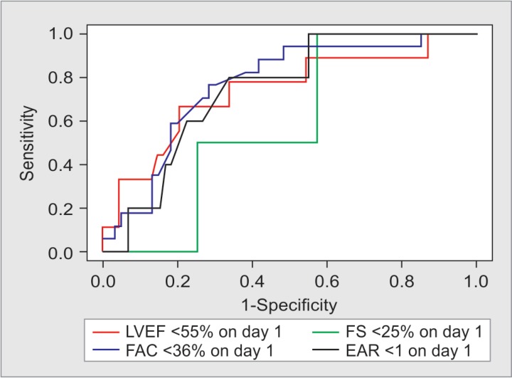
The ROC curves of CPK-MB for all the four echocardiographic parameters were constructed (Fig. 4); cutoff thresholds of CPK-MB and their corresponding sensitivity and specificity along with AUC with 95% CI are shown in Table 3. The AUCs of all, expect FS, are greater than 0.72.
Fig 4.
Receiver operating characteristics curves for CPK-MB as marker to predict echocardiographic evidence of myocardial dysfunction in patients with sepsis and septic shock
Table 3.
Receiver operating characteristics analysis for using CPK-MB as marker for echocardiographic parameters of myocardial dysfunctions on day 1 among all patients (n = 80)
| Echocardiographic parameters | Cutoffs for CPK-MB (IU/L) | Sensitivity | Specificity | AUC | 95% CI (DeLong method) |
|---|---|---|---|---|---|
| Left ventricular ejection fraction (<55%) | 152 | 0.79 | 0.67 | 0.74 | 0.54–0.93 |
| Fractional area change (<36%) | 105 | 0.72 | 0.76 | 0.76 | 0.64–0.88 |
| Fractional shortening (<25%) | 79 | 0.43 | 1.00 | 0.59 | 0.26–0.91 |
| E/A ratio (<1) | 119 | 0.66 | 0.80 | 0.74 | 0.57–0.92 |
AUC, area under curve; 95% CI, 95% confidence interval
DISCUSSION
Data from this study showed that CPK-MB at admission was significantly elevated in septic shock patients (both survivors and nonsurvivors) and was significantly higher compared to the NSS patients. Echocardiographic evidence of myocardial dysfunction was seen in 55% of SSSs; half having systolic dysfunction and 1/8th having diastolic dysfunction. None of the NSS patients had echocardiographic myocardial dysfunction. The CPK-MB as well as echocardiographic evidence of myocardial dysfunction improved over the next 7–10 days, and all patients had normal cardiac function at 1-month follow-up. Reduction in CPK-MB correlated well with improvement in PeLOD score, VIS, and echocardiographic parameters of myocardial dysfunction during the first 10 days.
Myocardial involvement in sepsis, the so-called “septic cardiomyopathy”,33 is well-documented but poorly understood. Fluid refractory septic shock patients may have low, normal, or high cardiac output. Hemodynamic parameters of cardiac output and systemic vascular resistance are reported to be heterogeneous and evolve over time in response to inotropic and vasopressor support.8,34 The incidence of myocardial dysfunction in pediatric septic shock varies from 50 to 80%,8–12,21,23,35 which maximizes by 1–3 days only to normalize by 7–10 days of illness,16,18 very similar to that we observed in the index study. The wide variation in the incidence of myocardial dysfunction may be due to the differences in patient selection (all septic shock or only fluid refractory septic shock, or after successful resuscitation as in the index study), delay between onset of illness and enrollment, use of different echocardiographic parameters and different cutoff values, choice of inotropes, and requirement for ventilation (cardiopulmonary interactions).
The incidence of diastolic dysfunction was lesser (12.5%) in the index study compared to that reported in literature. Raj et al. reported LV dysfunction in 53% of 30 patients; half of them had diastolic dysfunction.8 In an Indian study, myocardial dysfunction was reported in 71% of septic shock patients, diastolic being more common than systolic dysfunction (45.2 vs 32.3%).10 Another study from India revealed diastolic dysfunction in 41% of 56 children with fluid refractory septic shock. Mortality was higher in patients with diastolic dysfunction.9 It has been realized that diastolic dysfunction comes late and coincides with increasing severity of septic shock. Thus, limited diastolic filling in addition to impaired ventricular ejection is closely related to higher mortality.36 The lower incidence of diastolic dysfunction in our study is probably due to the inclusion of only SSSs.
Overall, LV dysfunction is found to be associated with increased severity of illness scores and use of inotropes; however, it is not associated with higher mortality.10,35 No difference was observed in myocardial dysfunction parameters between SSSs and nonsurvivors in the index study as well. It is probably because echocardiography was performed after hemodynamic stabilization with inotropic support which might have acted as an equalizer in the two groups. It is supported by the fact that the nonsurvivors had significantly higher VIS compared to the survivors on day 1. A biochemical marker such as CPK-MB may help overcome such limitation of echocardiography.
Injury to cardiac myocytes results in cell membrane damage and leads to the release of cardiac biomarkers (cTnT, cTnI, CPK-MB, and brain natriuretic peptide) into circulation.36 Although cTns generated lot of clinical interest,8–10,12,18,21,23 CPK-MB failed to generate much clinical interest in pediatric septic shock, as initial studies failed to help in predicting clinical outcome compared to cTns.18,23 Thus, its significance largely remained unexplored. In the index study also, though septic shock patients (survivors and nonsurvivors both) had markedly elevated CPK-MB compared to NSS patients, it failed to differentiate between survivors and nonsurvivors. Surprisingly, we found that subsequent fall of CPK-MB in SSSs paralleled to and was correlating well with improvement in clinical scores of illness severity (e.g., VIS, p = 0.04; PeLOD score, p < 0.01) and echocardiographic measures of myocardial dysfunction, both systolic (e.g., LVEF and FAC, p ≤ 0.01) and diastolic (EAR, p = 0.037) ones.
It is one of the largest longitudinal studies (n = 80) of myocardial dysfunction in pediatric sepsis and septic shock. To the best of our knowledge, it is the first pediatric study to document the longitudinal behavior of CPK-MB in septic shock. However, we are well aware of the limitations of including only survivors of septic shock. It would be interesting to know the longitudinal behavior of CPK-MB in patients who progressed from NSS to septic shock and among those who could not survive the catecholamine-resistant septic shock. Presence of multiorgan dysfunction syndrome in many of our patients might have erroneously affected the CPK-MB as the latter gets released from many other tissues in addition to the myocardium. There are other sources of CPK-MB in addition to the myocardium. Thus, his study was conducted during a period when the unit practice was to start dopamine, followed by dobutamine, maximizing them to 20 μg/kg/minute each and then to add adrenaline or noradrenaline in nonresponsive septic shock. Such practice led to high VIS in the studied patients by the time echocardiography was done. Such high VIS has a potential for underestimating the echocardiographic parameters. Half of the patients were ventilated in both the groups, creating further potential for underestimation due to cardiopulmonary interactions. Ideally, echocardiography should have been done in early stages of resuscitation (before starting inotropes and ventilation) by two independent trained pediatric cardiologists; however, even for research protocols, it is not feasible in resource-limited settings like ours. There is potential selection bias due to the inability to enroll consecutive patients. Despite associated limitations with one postgraduate pediatric trainee enrolling, consenting, performing serial echocardiography, and following up all patients, it is the reality of clinical research in the low- and low-middle income economies. Imputing the 10th day CPK-MB values of NSS patients for 1 month CPK-MB values for SSSs was a statistically pragmatic decision; however, it may not be without concerns. It also would have been interesting to correlate the increasing CPK-MB levels with the different underlying diseases and the level of bacteremia. It is a single-center study and findings are likely to be relevant in similar settings only. The information obtained may pave the way for further studies on a larger sample size with early echocardiographic examination by trained pediatric cardiologists to determine the prognostic and monitoring value of CPK-MB. It may be a useful marker for pediatric septic shock in low-/low-middle income economies and other resource-limited settings.
CONCLUSION
Echocardiographic evidence of myocardial dysfunction is common in septic shock despite being on high inotropic and ventilator support. With supportive management, it starts improving after day 3 and is completely reversible by day 10. The CPK-MB is significantly elevated among septic shock patients compared to the NSS patients. The initial CPK-MB levels were not predictive of mortality; however, its subsequent fall among SSSs paralleled with improvement in clinical status and myocardial functions. The CPK-MB isoenzyme may be an affordable and promising monitoring tool for septic cardiomyopathy in pediatric septic shock in resource-limited settings.
Footnotes
Source of support: Nil
Conflict of interest: None
REFERENCES
- 1.Singer M, Deutschman CS, Seymour CW, Shankar-Hari M, Annane D, Bauer M, et al. The third international consensus definitions for sepsis and septic shock (Sepsis-3). JAMA. 2016;315(8):801–810. doi: 10.1001/jama.2016.0287. DOI: [DOI] [PMC free article] [PubMed] [Google Scholar]
- 2.Mangia CMF, Kissoon N, Carcillo JA. Sepsis and septic shock: a global overview. J Pediatr Infect Dis. 2009;4(2):71–76. doi: 10.3233/JPI-2009-0157. DOI: [DOI] [Google Scholar]
- 3.Weiss SL, Fitzgerald JC, Pappachan J, Wheeler D, Jaramillo-Bustamante JC, Salloo A, et al. Global epidemiology of pediatric severe sepsis: the sepsis prevalence, outcomes, and therapies study. Am J Respir Crit Care Med. 2015;191(10):1147–1157. doi: 10.1164/rccm.201412-2323OC. DOI: [DOI] [PMC free article] [PubMed] [Google Scholar]
- 4.Sankar J, Ismail J, Sankar MJ, Suresh CP, Meena RS. Fluid bolus over 15-20 vs 5-10 minutes each in the first hour of resuscitation in children with septic shock: a randomized controlled trial. Pediatr Crit Care Med. 2017;18(10):e435–e445. doi: 10.1097/PCC.0000000000001269. DOI: [DOI] [PubMed] [Google Scholar]
- 5.Sankar J, Ismail J, Das R, Dev N, Chitkara A, Sankar MJ. Effect of severe vitamin D deficiency at admission on shock reversal in children with septic shock. J Intensive Care Med. 2019;34(5):397–403. doi: 10.1177/0885066617699802. DOI: [DOI] [PubMed] [Google Scholar]
- 6.Poddar B, Gurjar M, Singh S, Aggarwal A, Baronia A. Reduction in procalcitonin level and outcome in critically ill children with severe sepsis/septic shock-a pilot study. J Crit Care. 2016;36:230–233. doi: 10.1016/j.jcrc.2016.07.022. DOI: [DOI] [PubMed] [Google Scholar]
- 7.Ghosh S, Baranwal AK, Bhatia P, Nallasamy K. Suspecting hyperferritinemic sepsis in iron-deficient population: do we need a lower plasma ferritin threshold? Pediatr Crit Care Med. 2018;19(7):e367–e373. doi: 10.1097/PCC.0000000000001584. DOI: [DOI] [PubMed] [Google Scholar]
- 8.Raj S, Killinger JS, Gonzalez JA, Lopez L. Myocardial dysfunction in pediatric septic shock. J Pediatr. 2014;164(1):72–77. doi: 10.1016/j.jpeds.2013.09.027. DOI: [DOI] [PubMed] [Google Scholar]
- 9.Sankar J, Das RR, Jain A, Dewangan S, Khilnani P, Yadav D, et al. Prevalence and outcome of diastolic dysfunction in children with fluid refractory septic shock--a prospective observational study. Pediatr Crit Care Med. 2014;15(9):e370–e378. doi: 10.1097/PCC.0000000000000249. DOI: [DOI] [PubMed] [Google Scholar]
- 10.Jain A, Sankar J, Anubhuti A, Yadav DK, Sankar MJ. Prevalence and outcome of sepsis-induced myocardial dysfunction in children with ‘Sepsis’ ‘With’ and ‘Without Shock’-a prospective observational study. J Trop Pediatr. 2018;64(6):501–509. doi: 10.1093/tropej/fmx105. DOI: [DOI] [PubMed] [Google Scholar]
- 11.Ranjit S, Kissoon N. Bedside echocardiography is useful in assessing children with fluid and inotrope resistant septic shock. Indian J Crit Care Med. 2013;17(4):224–230. doi: 10.4103/0972-5229.118426. DOI: [DOI] [PMC free article] [PubMed] [Google Scholar]
- 12.El-Zayat RS, Shalaby AG. Mitral annular plane systolic excursion as a predictor of mortality in children with septic shock. Pediatr Crit Care Med. 2018;19(9):e486–e494. doi: 10.1097/PCC.0000000000001661. DOI: [DOI] [PubMed] [Google Scholar]
- 13.Kumar A, Thota V, Dee L, Olson J, Uretz E, Parrillo JE. Tumor necrosis factor alpha and interleukin 1beta are responsible for in vitro myocardial cell depression induced by human septic shock serum. J Exp Med. 1996;183(3):949–958. doi: 10.1084/jem.183.3.949. DOI: [DOI] [PMC free article] [PubMed] [Google Scholar]
- 14.Thiru Y, Pathan N, Bignall S, Habibi P, Levin M. A myocardial cytotoxic process is involved in the cardiac dysfunction of meningococcal septic shock. Crit Care Med. 2000;28(8):2979–2983. doi: 10.1097/00003246-200008000-00049. DOI: [DOI] [PubMed] [Google Scholar]
- 15.Turner A, Tsamitros M, Bellomo R. Myocardial cell injury in septic shock. Crit Care Med. 1999;27(9):1775–1780. doi: 10.1097/00003246-199909000-00012. DOI: [DOI] [PubMed] [Google Scholar]
- 16.Bouhemad B, Nicolas-Robin A, Arbelot C, Arthaud M, Féger F, Rouby J-J. Isolated and reversible impairment of ventricular relaxation in patients with septic shock. Crit Care Med. 2008;36(3):766–774. doi: 10.1097/CCM.0B013E31816596BC. DOI: [DOI] [PubMed] [Google Scholar]
- 17.Pulido JN, Afessa B, Masaki M, Yuasa T, Gillespie S, Herasevich V, et al. Clinical spectrum, frequency, and significance of myocardial dysfunction in severe sepsis and septic shock. Mayo Clin Proc. 2012;87(7):620–628. doi: 10.1016/j.mayocp.2012.01.018. DOI: [DOI] [PMC free article] [PubMed] [Google Scholar]
- 18.Oliveira NS, Silva VR, Castelo JS, Elias-Neto J, Pereira FEL, Carvalho WB. Serum level of cardiac troponin I in pediatric patients with sepsis or septic shock. Pediatr Crit Care Med. 2008;9(4):414–417. doi: 10.1097/PCC.0b013e31817e2b33. DOI: [DOI] [PubMed] [Google Scholar]
- 19.Yu Y-J, Su A-H, Yang H-B, Chen J-X. Intermedin1-53 protects cardiac function in rats with septic shock via inhibiting oxidative stress and cardiomyocyte apoptosis. Eur Rev Med Pharmacol Sci. 2018;22(9):2906–2913. doi: 10.26355/eurrev_201805_14993. DOI: [DOI] [PubMed] [Google Scholar]
- 20.El-Awady MS, Nader MA, Sharawy MH. The inhibition of inducible nitric oxide synthase and oxidative stress by agmatine attenuates vascular dysfunction in rat acute endotoxemic model. Environ Toxicol Pharmacol. 2017;55:74–80. doi: 10.1016/j.etap.2017.08.009. DOI: [DOI] [PubMed] [Google Scholar]
- 21.Fenton KE, Sable CA, Bell MJ, Patel KM, Berger JT. Increases in serum levels of troponin I are associated with cardiac dysfunction and disease severity in pediatric patients with septic shock. Pediatr Crit Care Med. 2004;5(6):533–538. doi: 10.1097/01.PCC.0000144711.97646.0C. DOI: [DOI] [PubMed] [Google Scholar]
- 22.Basu S, Frank LH, Fenton KE, Sable CA, Levy RJ, Berger JT. Two-dimensional speckle tracking imaging detects impaired myocardial performance in children with septic shock, not recognized by conventional echocardiography. Pediatr Crit Care Med. 2012;13(3):259–264. doi: 10.1097/PCC.0b013e3182288445. DOI: [DOI] [PubMed] [Google Scholar]
- 23.Lodha R, Arun S, Vivekanandhan S, Kohli U, Kabra SK. Myocardial cell injury is common in children with septic shock. Acta Paediatr. 2009;98(3):478–481. doi: 10.1111/j.1651-2227.2008.01095.x. DOI: [DOI] [PubMed] [Google Scholar]
- 24.Goldstein B, Giroir B, Randolph A. International pediatric sepsis consensus conference: definitions for sepsis and organ dysfunction in pediatrics. Pediatr Crit Care Med. 2005;6(1):2–8. doi: 10.1097/01.PCC.0000149131.72248.E6. DOI: [DOI] [PubMed] [Google Scholar]
- 25.Dellinger RP, Levy MM, Carlet JM, Bion J, Parker MM, Jaeschke R, et al. Surviving Sepsis Campaign: international guidelines for management of severe sepsis and septic shock: 2008. Intensive Care Med. 2008;34(1):17–60. doi: 10.1007/s00134-007-0934-2. DOI: [DOI] [PMC free article] [PubMed] [Google Scholar]
- 26.Kleinman ME, Chameides L, Schexnayder SM, Samson RA, Hazinski MF, Atkins DL, et al. Part 14: pediatric advanced life support: 2010 American Heart Association Guidelines for Cardiopulmonary Resuscitation and Emergency Cardiovascular Care. Circulation. 2010;122(18 Suppl 3:):S876–S908. doi: 10.1161/CIRCULATIONAHA.110.971101. DOI: [DOI] [PubMed] [Google Scholar]
- 27.Gaies MG, Gurney JG, Yen AH, Napoli ML, Gajarski RJ, Ohye RG, et al. Vasoactive-inotropic score as a predictor of morbidity and mortality in infants after cardiopulmonary bypass. Pediatr Crit Care Med. 2010;11(2):234–238. doi: 10.1097/PCC.0b013e3181b806fc. DOI: [DOI] [PubMed] [Google Scholar]
- 28.R Core Team. R: A language and environment for statistical computing. R Foundation for Statistical Computing. Austria.: Jun 30, 2018. 2018. available at. , accessed at. [Google Scholar]
- 29.Wickham H. ggplot2: Elegant Graphics for Data Analysis. New York: Springer-Verlag; Jun 30, 2018. 2016, available at. , accessed at. [Google Scholar]
- 30.Fox J, Weisberg S. An {R} Companion to Applied Regression, 2nd ed. Thousand Oaks CA: Sage; Jun 30, 2018. 2011, available at. , accessed at. [Google Scholar]
- 31.Robin X, Turck N, Hainard A, Tiberti N, Lisacek F, Sanchez J-C, et al. pROC: an open-source package for R and S+ to analyze and compare ROC curves. BMC Bioinformatics. 2011;12:77. doi: 10.1186/1471-2105-12-77. DOI: [DOI] [PMC free article] [PubMed] [Google Scholar]
- 32.Trapletti A, Hornik K,. tseries: Time Series Analysis and Computational Finance. R package version 0.10-45. https://CRAN.R-project.org/package=tseries Jun 30, 2018. https://CRAN.R-project.org/package=tseries 2018, available at. , accessed at.
- 33.Hawiger J. Heartfelt sepsis: microvascular injury due to genomic storm. Kardiol Pol. 2018;76(8):1203–1216. doi: 10.5603/KP.a2018.0146. DOI: [DOI] [PubMed] [Google Scholar]
- 34.Ceneviva G, Paschall JA, Maffei F, Carcillo JA. Hemodynamic support in fluid-refractory pediatric septic shock. Pediatrics. 1998;102(82):e19. doi: 10.1542/peds.102.2.e19. DOI: [DOI] [PubMed] [Google Scholar]
- 35.Williams FZ, Sachdeva R, Travers CD, Walson KH, Hebbar KB. Characterization of myocardial dysfunction in fluid- and catecholamine-refractory pediatric septic shock and its clinical significance. J Intensive Care Med. 2019;34(1):17–25. doi: 10.1177/0885066616685247. DOI: [DOI] [PubMed] [Google Scholar]
- 36.Walley KR. Sepsis-induced myocardial dysfunction. Curr Opin Crit Care. 2018;24(4):292–299. doi: 10.1097/MCC.0000000000000507. DOI: [DOI] [PubMed] [Google Scholar]


