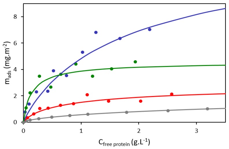
Figure 2.
Adsorption isotherms of yeast protein extract (S288c) adsorbed on different silica nanoparticles in Dulbecco’s phosphate-buffered saline (DPBS) buffer (pH 7.4). The curves depict isotherms fitted by the Langmuir–Freundlich model for the following silica nanoparticles: S10 (red); S30 (blue); S80 (green); and polydisperse nanoparticles (NPs) (grey) taken from Mathé et al. [34]. The dots are experimental points associated to a single isotherm.

