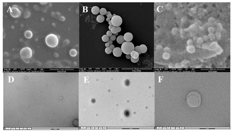
Figure 2.
SEM photographs of liposome (A), invasome (B), and transfersome formulations (C), and TEM photographs of liposome (D), invasome (E), and transfersome formulations (F). Reprinted with permission from reference [33]. Copyright 2018, Elsevier.

