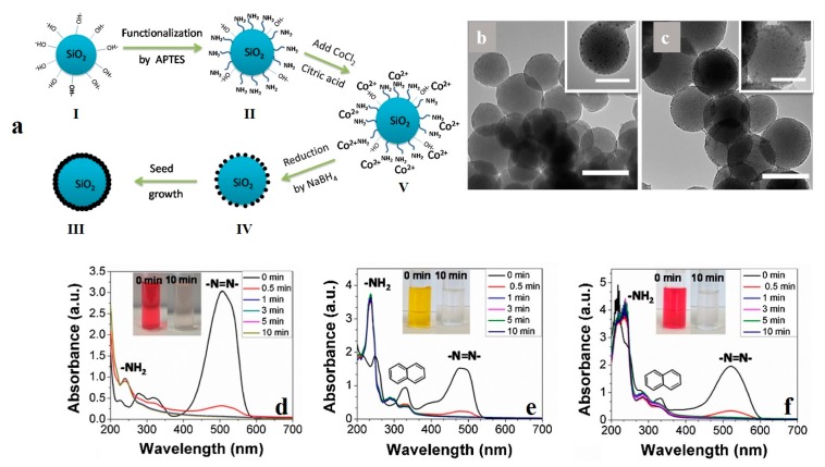
Figure 5.
(a) The synthesis procedure for SiO2-Co core-shell nanoparticles. TEM images imply the SiO2-Co core-shell nanoparticles prepared from two different Co2+ precursor amounts of (b) 1 mM and (c) 2 mM Co2+ (Scale bar in the main image and inset represents 100 nm and 50 nm, respectively). UV/Vis spectra indicate the model dye degradation by the core-shell nanoparticles over time (note that the solutions’ pH was acidic (pH 2.5) and the initial dye concentration was 0.076 mM) (d) Methyl Orange. (e) Orange G. (f) Amaranth. Reproduced with permission [103]. Copyright 2016, Elsevier.

