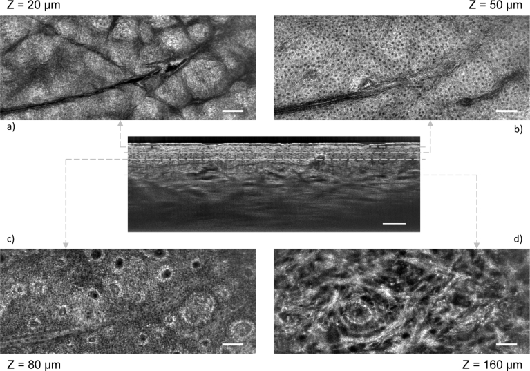
Fig. 3.
LC-OCT C-scan images of healthy human skin in vivo (back of the hand), obtained for several layers of the skin: a) stratum corneum, b) stratum spinosum, c) stratum basale and d) papillary dermis. The depth (Z) of each layers is indicated, counted from the surface of the skin. The C-scans are correlated to a B-scan obtained by switching from C-scan mode to B-scan mode. Scale bars: 100 µm.

