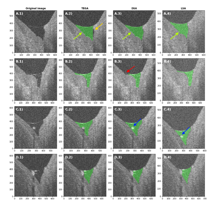
Fig. 5.
Comparison between the TBSA (column 2), DSA (3) and LSA (4) in rare cases of challenging segmentation tasks (1). None of the images were part of the training dataset. (A) Not segmented lateral cavity (orange arrow), (B) irregular meniscus shape, (C) small debris and (D) larger debris cutting the tear meniscus area in two parts. Green arrows indicate example areas where the bordering pixels are included in or excluded from the tear meniscus area. The red arrow indicates a non-segmented region of the tear meniscus area. The blue arrows indicate holes in the segmented area, where debris is present. Images in columns 1-3 are cropped from a image, while images in column 4 are cropped from a image. The axes represent µm in tear fluid.

