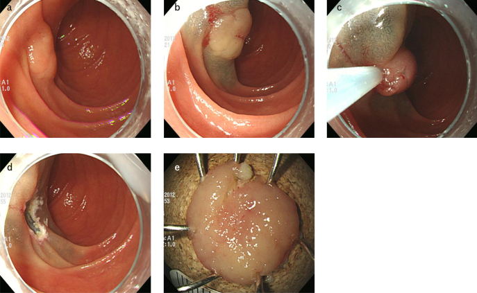
Figure 2. a–e.
Endoscopic mucosal resection. (a) A slightly elevated lesion is observed at the second portion of the duodenum. (b) A saline solution containing small amounts of epinephrine and indigo carmine dye is injected beneath the lesion to elevate the lesion. (c) A snare resection is performed using a blended electrosurgical current. (d) The lesion is completely removed. (e) The resected specimen.

