
Figure 1. Regulation of serum urate.
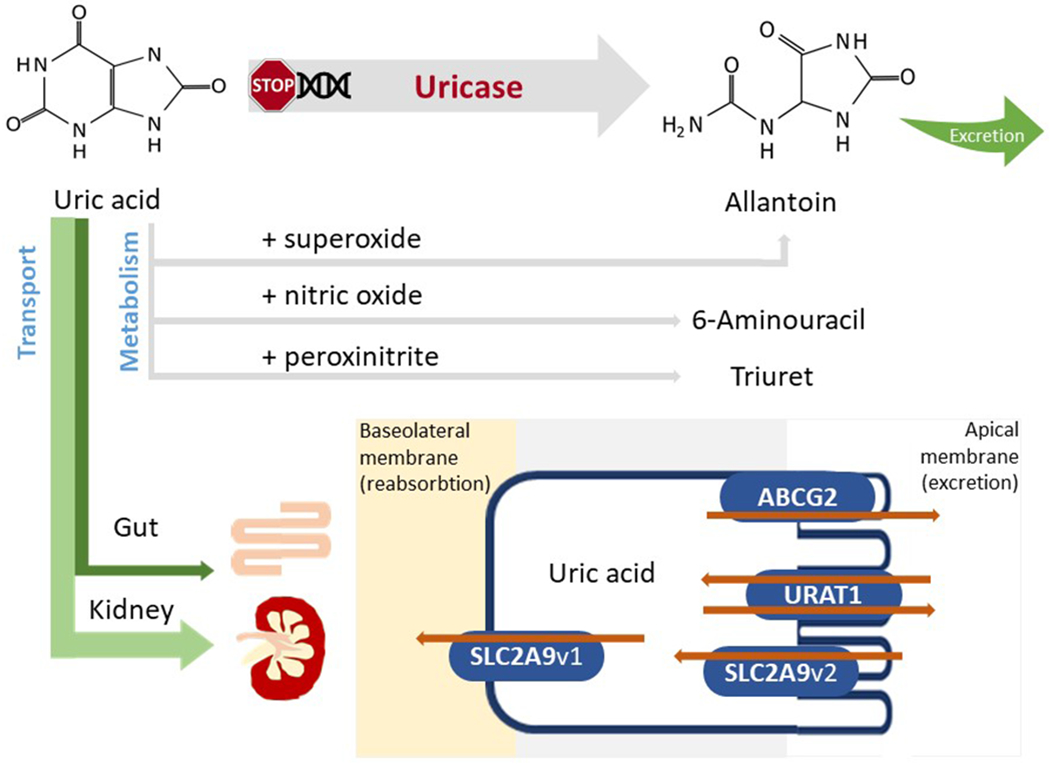
Following sequential mutations in the uricase gene, humans and higher primates do not express functional uricase, needed to metabolize uric acid to allantoin. Serum urate can interact with reactive species generating substances or radicals such as allantoin, 6-aminouracil or triuret. However, most of the serum urate will be excreted via the kidney or, alternatively, via the gut. Both proximal tubule and intestinal cells express several urate transporters (collectively known as the transportasome) that are responsible for the excretion and/or reabsorption of urate. Several main urate transporters are illustrated: URAT1, GLUT9, OAT4 and OAT10 are involved in urate reabsorption at the level of the apical membrane (brush border) of proximal renal tubular cells; GLUT9 is responsible for urate transport out of the cell via the basolateral membrane into the blood; ABCG2 is a unidirectional transporter mediating the secretion of urate via the apical membrane; OAT1 and OAT3 localized on the basolateral membrane are involved in urate excretion. OAT, organic anion transporter; URAT1, urate transporter 1; GLUT9, glucose transporter 9; ABCG2, ATP-binding cassette super-family G member 2.