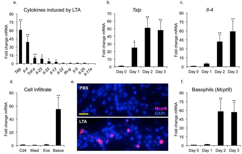
Fig 1. A Tslp-basophil-Il4 axis is initiated by intradermal LTA. (a) Mice were injected ID with PBS or LTA.
Skin samples at site of injection were harvested 48 hours later and mRNA was analyzed by rt-PCR for expression of cytokine genes. (b) Tslp and (c) Il-4 gene expression were analyzed by RT-PCR in mice injected ID with LTA and harvested at the indicated time points post treatment. (d) Mice injected ID with LTA were analyzed 48 hours later by RT-PCR for the expression of cell infiltrates. Quantitation by RT-PCR for the markers cd4 (T cells), kit (mast cells), mbp (eosinophils) and mcpt8 (basophils). (e) Staining of skin shows increased Mcpt8 expression in the dermis upon LTA treatment. Bar = 40 mm. (f) Mice injected ID with LTA were harvested at the indicated time points post treatment and analyzed by RT-PCR for expression of the basophil marker (mcpt8). Data are mean, ± SEM, n = 6. *P<0.05; **P<0.01 compared to control.

