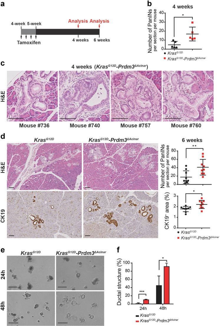
Fig. 2. Loss of prdm3 promotes acinar-to-ductal metaplasia and PanIN lesions formation.
a Ptf1aCreER;KrasG12D and Ptf1aCreER;KrasG12D;Prdm3flox/flox mice at 4–5 weeks of age were injected 4 times on alternating days with tamoxifen. Recombined mice were analyzed at 4 and 6 weeks post tamoxifen injection. b The number of PanINs per section for KrasG12D (n = 5) vs. KrasG12D-Prdm3ΔAcinar (n = 5) mice at 4 weeks post tamoxifen injection. c Representative images of high-grade PanINs in KrasG12D-Prdm3ΔAcinar mice at 4 weeks after tamoxifen injection. d Hematoxylin-eosin staining (H&E) and immunohistochemistry staining for the ductal marker Cytokeratin 19 (CK19). Quantification of the number of PanINs, as well as the percent of pancreatic area that is CK19+ in KrasG12D (n = 9) vs. KrasG12D-Prdm3ΔAcinar (n = 9) mice 6 weeks post-tamoxifen injection. e Images of acinar cell explants embedded in Matrigel at 24 and 48 h. f Quantification of the percent of ductal-like structures in explants derived from KrasG12D (n = 3) and KrasG12D-Prdm3ΔAcinar (n = 3) mice at 4 weeks post-tamoxifen injection. Data show mean ± SD. Statistical analysis: Two-tailed t-test. *p < 0.05, **p < 0.01, ***p < 0.001. Scale: 100 μm.

