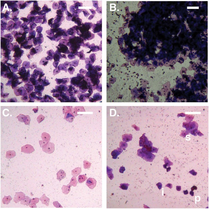
Figure 1.
Bright-field micrographs (100X total magnification). (A) No Amsel-BV sample with cell count >100/100X field, showing predominantly superficial cells shed as singletons. (B) Symptomatic Amsel-BV sample with cell count >100/100X field, showing predominantly superficial cells shed in large aggregates. (C) No Amsel-BV sample with cell count <50/100X field, showing predominantly superficial cells. (D) Asymptomatic Amsel-BV sample with cell count <50/100X, showing superficial cells (example marked s) shed in combination with intermediate (example marked i) and parabasal (example marked p) cells. Scale bars represent 100 μm.

