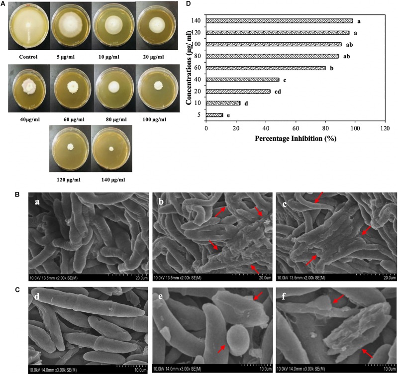
FIGURE 9.
(A) Plates showing antifungal activity of Melia leaf extract (MLE)-silver nanoparticles (AgNPs) at different concentrations (5, 10, 20, 60, 80, 100, 120, and 140 μg/ml) after 7 days of incubation at 28°C. (B) Scanning electron microscope (SEM) images of Fusarium oxysporum hyphae in the presence of water (a), 100 μg/ml (b) and 120 μg/ml of MLE-AgNPs (c). (C) SEM images of conidia of F. oxysporum with sterile water (d) and MLE-AgNPs (e,f) for 24 h. (D) Effect of different concentrations (5, 10, 20, 40, 60, 80, 100, 120, and 140 μg/ml) of MLE-AgNPs after 7 days on F. oxysporum by calculating percentage inhibition (%). Vertical bars represent standard error between various replicates of the same treatments. Values with the same letter differ non-significantly (P ≥ 0.05) as created by ANOVA and Duncan’s new multiple range test.

