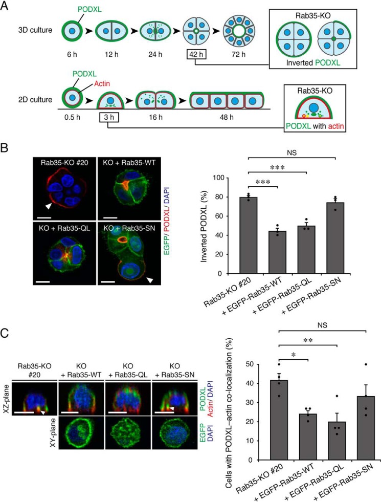
Figure 1.
Active Rab35 is required for PODXL trafficking under 2D and 3D culture conditions. A, schematic representation of PODXL localization during 3D cyst and 2D monolayer formation of MDCK II cells (modified from Ref. 6). In a 3D Rab35-KO cell culture, PODXL (green) remained on the outer membrane at 42 h after plating (Inverted PODXL) (top row, 3D culture). In a 2D Rab35-KO cell culture, endocytosed PODXL from the membrane attaching to the bottom plane of the dish localized and accumulated on actin-rich structures (red) at 3 h after plating (PODXL with actin) (bottom row, 2D culture). B, Rab35-KO (#20) and its rescued cells (+EGFP-Rab35 (WT, QL, or SN; shown in green)) were plated on Matrigel and fixed at 42 h after plating. The cells were stained for PODXL (red) and DAPI (blue), followed by counting of the inverted PODXL (30 cysts/condition). The arrowheads show PODXL localized on the outer membrane. Scale bars, 10 μm. The graph shows the means and S.E. (error bars) of three independent experiments. ***, p < 0.001; NS, not significant (Dunnett's test). C, Rab35-KO (#20) and its rescued cells (+EGFP-Rab35 (WT, QL, or SN)) were plated on glass-bottom dishes and fixed at 3 h after plating. The cells were stained for PODXL (green), actin (red), and DAPI (blue), followed by counting of cells with co-localized PODXL and actin (30 cells/condition). The confocal xz-plane (top) and the xy-plane (bottom) are shown. The arrowheads show PODXL co-localizing with actin. Scale bars, 10 μm. The graph shows the means and S.E. (error bars) of four independent experiments. *, p < 0.05; **, p < 0.01; NS, not significant (Dunnett's test).

