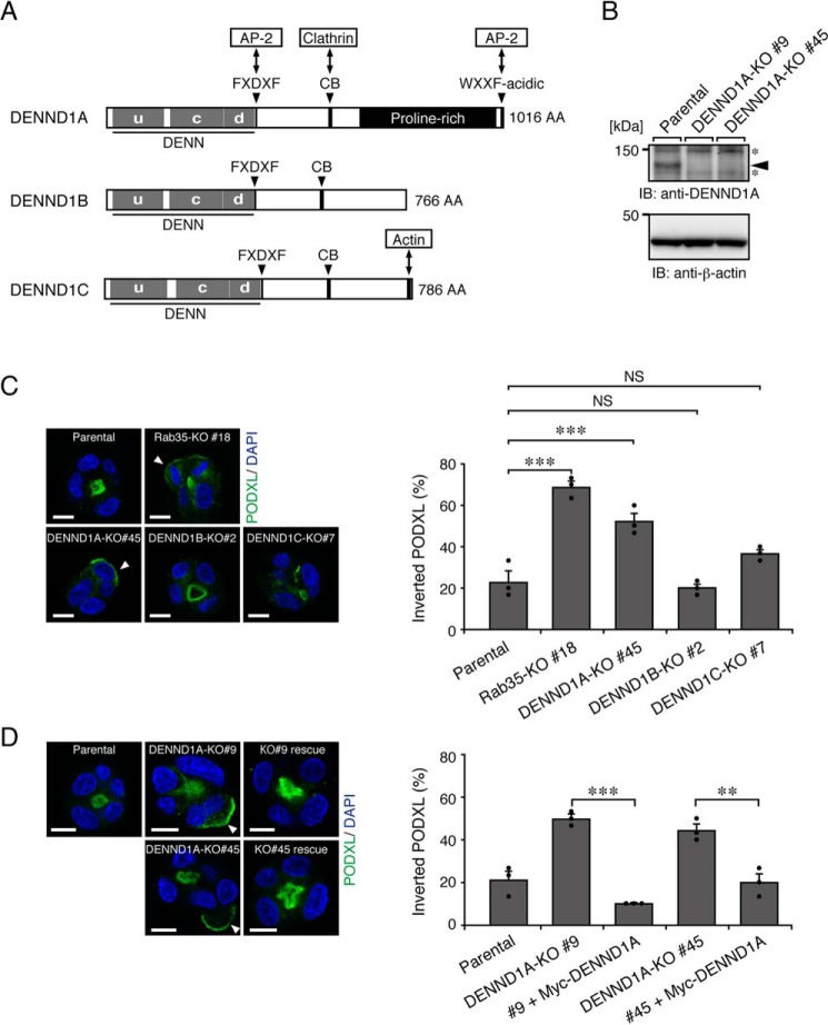
Figure 2.
DENND1A-KO, but not DENND1B-KO or DENND1C-KO, induces the inverted localization of PODXL in 3D cysts. A, schematic representation of mouse DENND1 family proteins. The domains and motifs are depicted according to previous reports (15, 17) and the UniProtKB (DENND1A, Q8K382; DENND1B, Q3U1T9; DENND1C, Q8CFK6). uDENN, upstream DENN; cDENN, central/core DENN; dDENN, downstream DENN; FXDXF, AP-2 α-ear platform subdomain–binding motif; CB, clathrin heavy chain–binding motif; WXXF-acidic, AP-2 α-ear sandwich subdomain–binding motif; AA, amino acids. B, lysates of parental cells and two DENND1A-KO cells (#9 and #45) were analyzed by immunoblotting (IB) with anti-DENND1A and anti-β-actin antibodies. The arrowhead indicates the position of endogenous DENND1A. The asterisks indicate nonspecific bands of the primary antibody. C, parental, Rab35-KO, DENND1A-KO, DENND1B-KO, and DENND1C-KO cells were plated on Matrigel and fixed at 42 h after plating. The cells were stained for PODXL (green) and DAPI (blue), followed by counting of the inverted PODXL (30 cysts/condition). The arrowheads show PODXL localizing on the outer membrane. Scale bars, 10 μm. The graph shows the means and S.E. (error bars) of three independent experiments. ***, p < 0.001; NS, not significant (Dunnett's test). D, parental cells, DENND1A-KO (#9 and #45) cells, and their rescued cells (+Myc-DENND1A) were plated on Matrigel and fixed at 42 h after plating, followed by counting of the inverted PODXL (30 cysts/condition). The arrowheads show PODXL localizing on the outer membrane. Scale bars, 10 μm. The graph shows the means and S.E.(error bars) of three independent experiments. ***, p < 0.001; **, p < 0.01 (Tukey's test).

