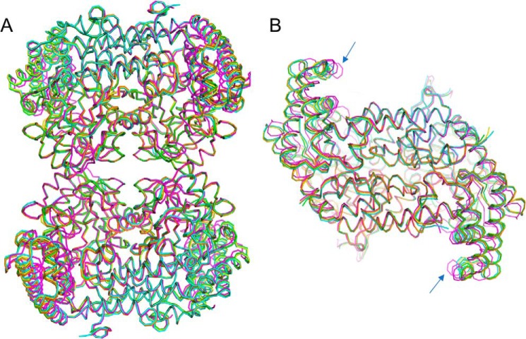
Figure 3.
Structural similarity of the SHMT8 structures in this study. Structures are colored as follows: Essex SHMT8 in complex with PLP (yellow), PLP-Gly (blue), PLP-Gly with THF (magenta), and Forrest SHMT8 in complex with PLP (green), and PLP-Gly (orange). A, a superposition of the polypeptide backbone the five tetramers. B, top-down view of the five obligate dimers, showing the slight difference (blue arrows) in the orientation of the small domain in the ternary complex with PLP-Gly and FTHF (magenta). A similar conformational change of the small domain was observed in the murine SHMT complex with FTHF (27).

