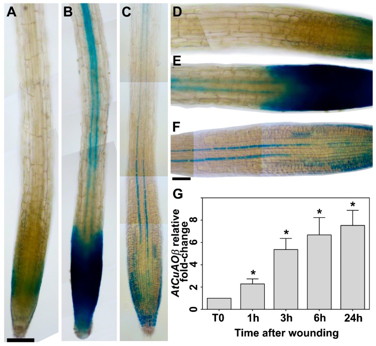
Figure 1.
Analysis of AtCuAOβ gene expression upon leaf wounding by GUS staining analysis (root) and RT-qPCR (whole seedlings). (a–f) Light microscopy analysis after GUS staining of roots from 7-day-old AtCuAOβ-promoter::GFP-GUS seedlings unwounded (a–f) or leaf-wounded after 3 h from the injury (b,e). The staining reaction was allowed to proceed for 2 h (a,d unwounded, and b,e wounded leaf) or overnight (c,f; unwounded). Shown images were obtained aligning serial overlapping micrographs of the same root using Photoshop Software (Adobe). Ten plants from three independent experiments were analyzed and images from a single representative experiment is shown. Bar: 200 μm (a–c); 100 μm (d–f). (g) RT-qPCR analysis in 7-day-old wild type (WT) whole seedlings at 0, 1, 3, 6 and 24 h time-points from cotyledonary leaf injury. Three biological replicates each with three technical replicates were performed (n = 3). AtCuAOβ mRNA level after wounding is relative to that of the corresponding unwounded plant for each time point. The significance levels (p-values) between the relative mRNA level at each time and the mRNA level of control unwounded plant at time 0 (T0), which is assumed to be one, have been calculated with one-way analysis of variance (ANOVA) followed by Sidak’s multiple comparison test (*; p-values ≤ 0.05).

