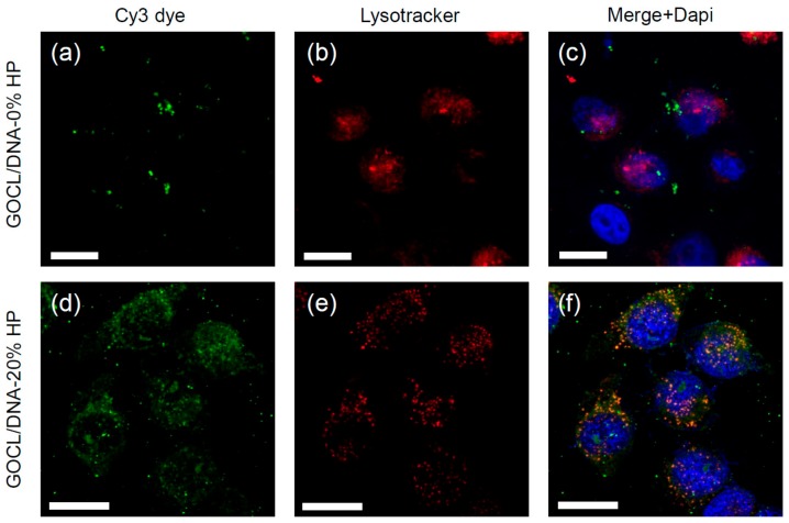
Figure 5.
Representative confocal microscopy images of of MDA-MB cells treated with fluorescently labelled (green) pristine (panels a–c) and biocoronated grapholipoplexes (HP = 20%) (panels d–f). Cells were stained with LysoTracker Deep Red (red), a well-known lysosomal marker, and Dapi to stain the nuclei. Scale bars are 5 microns.

