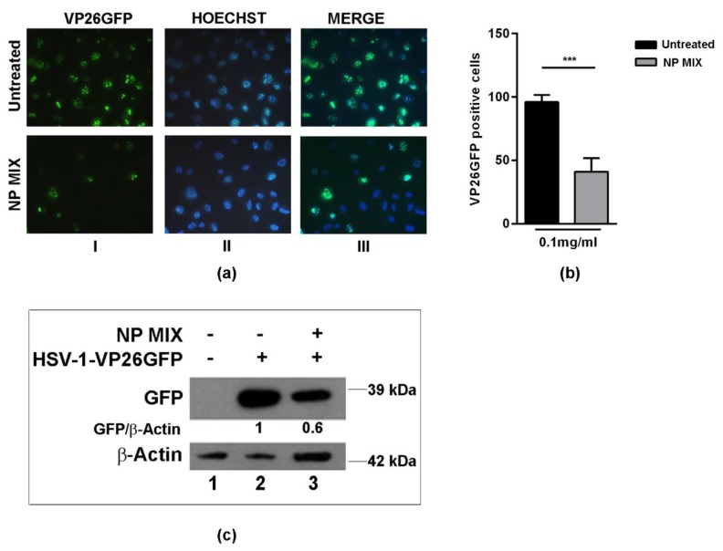
Figure 5.
Effect of the NP MIX treatment on HSV-1 replication. Vero cells were either infected or mock-infected with HSV-1-VP26 GFP (green fluorescent protein-tagged capsid protein VP26) at multiplicity of infection (MOI) 1, as described in the Materials and Methods section. Then, the cells were analysed at 24 h post infection (p.i.): (a) fluorescent images showed the green dots representing the VP26 GFP viral antigen localization (I)—Hoechst (blue) was used to stain the nuclei (II) and the merged images are shown in column III; (b) the graph is indicative of the percentage of VP26 GFP-positive cells; (c) Western blot analysis of VP26 GFP-tagged protein. Data are expressed as a mean (± SD) of at least three experiments, and asterisks (***) indicate the significance of p-values less than 0.001.

