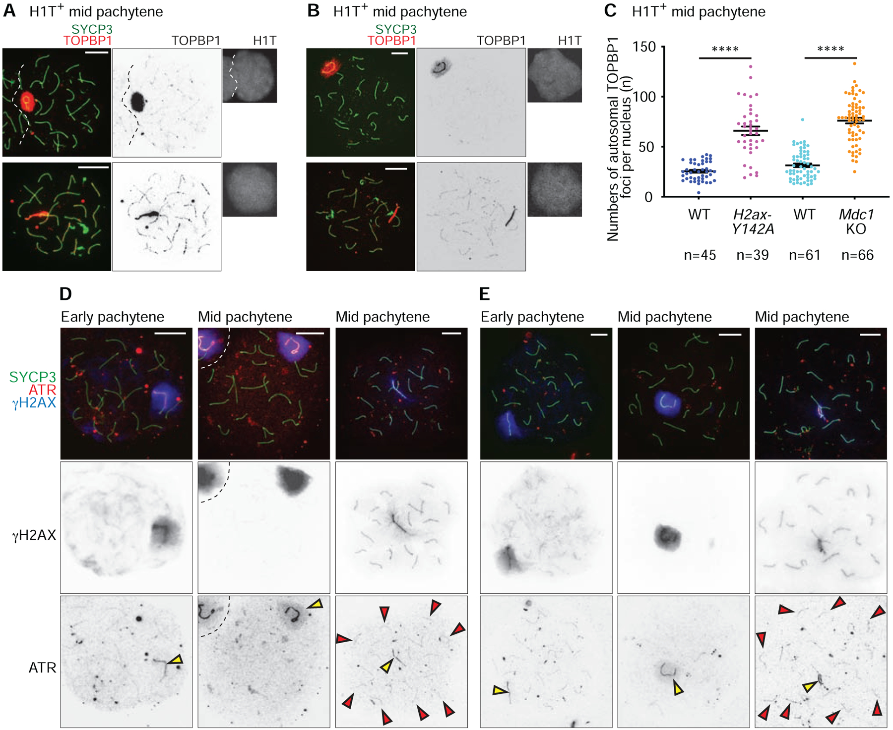
Figure 5. DDR factors centered on ATR signaling are sequestered from autosomes to the sex chromosomes at the onset of MSCI.

(A, B, D, E) Chromosome spreads of wild-type (WT) littermate control and H2ax-Y142A (A, D) or Mdc1KO (B, E) mid pachytene spermatocytes immunostained with antibodies raised against SYCP3 (A, B, D, E), TOPBP1 (A, B), H1T (A, B), γH2AX (D, E), and ATR (D, E). Yellow arrowheads indicate the sex chromosomes, and red arrowheads indicate ATR foci that persist on H2ax-Y142A autosomes (D, E). Scale bars: 10 μm.
(C) Numbers of TOPBP1 foci on autosomes in mid pachytene (H1T-positive) spermatocytes, shown as mean ± s.e.m. for 3 independent H2ax-Y142A littermate pairs (left) and 3 independent Mdc1KO littermate pairs (right). Total numbers of analyzed nuclei are indicated in the panels. **** p < 0.0001, Mann-Whitney U test.
See also Figure S5.
