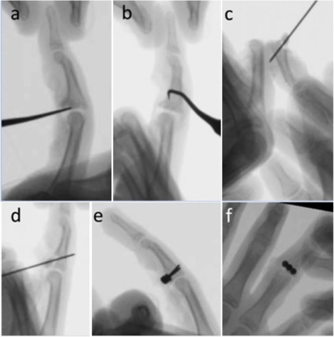
Figure 2.
Intraoperative fluoroscopy. (a) Dental pick is used to reduce impacted articular fragments and (b) free any formation of consolidated tissue. (c) The joint is “shotgunned” open exposing the joint allowing for (d) Kirschner-wire to reduce the fracture. (e) Lateral and (f) posteroanterior fluoroscopy of 3-hole plate with 3 nonlocking screws achieving concentric reduction of joint.

