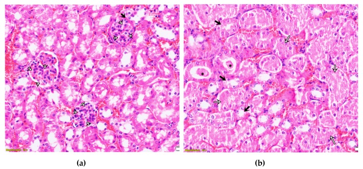
Figure A3.
Histopathology of the kidney of BALB/c mice receiving LysM lectin fraction: (a) Glomerular morphology disruptions. White arrows show vacuolated cells in glomeruli, the black arrow indicates Bauman’s space filled with proteinaceous fluid infiltration; (b) tubular morphology disruption. The black asterisk shows tubular protein cylinders; black arrows indicate distal tubular necrosis, white arrows indicate proximal tubular necrosis.

