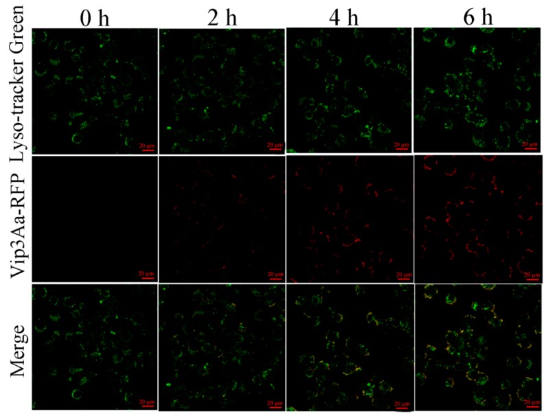
Figure 2.
Co-localization of Vip3Aa and lysosomes in Sf9 cells. Cells were treated with Vip3Aa-RFP for 0, 2, 4, and 6 h, respectively, and were stained with fluorescent probe LysoSensor™ Green DND-189 at 28 ℃ for 45 min. Then, the cells were observed under a confocal laser scanning microscope. Scale bar, 20 µm.

