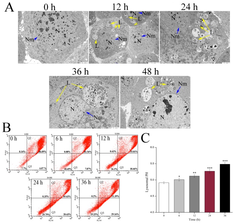
Figure 5.
Effects of Vip3Aa on lysosomes in Sf9 cells. (A) Representative photographs of lysosomes ultrastructure in Sf9 cells after exposure to Vip3Aa, obtained by TEM. N, nucleus. Nm, nuclear membrane (blue arrows). L, lysosomes (yellow arrows). Magnification, 0 h, 24 h, 36 h, and 48 h, 10000 ×; 12 h, 5000 ×. (B) The physicochemical property of lysosomes was detected by acridine orange (AO) staining in Sf9 cells. (C) The lysosomal pH in Sf9 cells was detected using LysoSensor Yellow/Blue DND-160. Significant tests from the corresponding controls (without Vip3Aa treatment) are indicated by * p < 0.05, ** p < 0.01, and *** p < 0.001.

