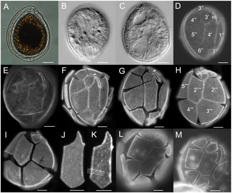
Figure 2.
Light (A–C) and epifluorescence (D–M) micrographs of cells of O. cf. ovata strain UNR-10 from Rio Grande do Norte, Brazil (A–K), and strain VGO614 from Madeira Island, Portugal (L–M): (A) broadly oval cell shape; (B–C) elongated chloroplasts and the posterior nucleus are visible; (D–G) apical view, note the variable size of the suture between plates 3′ and 5” and variable shape of plate 4′, plate 2′ extends between plates 2” and 3′ (F–G, arrow heads); (H–I) antapical view; (J–K) plate 4′ variable shape; (L–M) apical view of two cells from strain VGO614. Scale bars: 10 µm.

