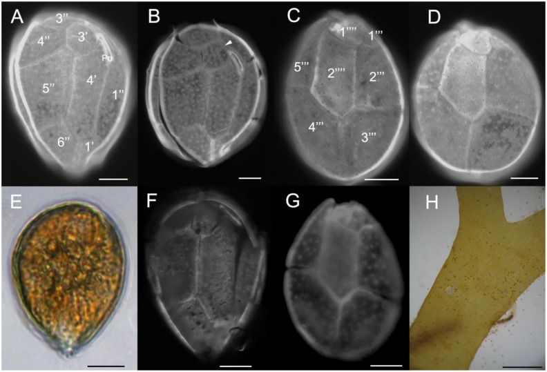
Figure 3.
Epifluorescence (A–D, F–G) and light (E, H) micrographs of cells of O. cf. ovata strain UNR-05 from Rio de Janeiro, Brazil (A–E); field cells from Forte, Bahia (F–G); and from a bloom at Rio de Janeiro (H): (A–B, F) apical view; (C–D, G) antapical view; (E) live cell; (H) field live cells associated to the macroalgae Canistrocarpus cervicornis during a bloom at Arraial do Cabo, Rio de Janeiro, Brazil. Scale bars: (A–G): 10 µm; H: 1000 µm.

