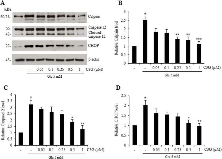
Fig. 4.
Protective effect of C3G against glutamate-induced ER stress apoptotic protein expression in HT22 cells. Cells were pretreated with C3G (0–1 μM) for 24 h, followed by 5 mM glutamate for 18 h. After treatment, (a) the level of calpain, caspase-12 and CHOP were determined by Western blot analysis, and β-actin was served as the loading control. (b, c, d) Relative protein levels were quantified by densitometry and the mean data from independent experiments were normalized to the results. The data represent the means of four independent samples ± SD. #p < 0.05 versus non-treated control, *p < 0.05, **p < 0.01, ***p < 0.001 versus 5 mM glutamate-treated cells

