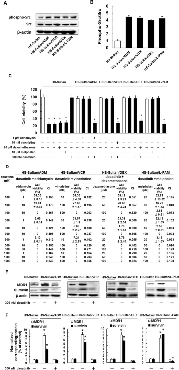
Fig. 4.

The src inhibitor dasatinib reversed drug resistance in HS-Sultan/ADM, HS-Sultan/VCR, HS-Sultan/DEX, and HS-Sultan/L-PAM cells. a Phosphorylated Src expression levels were assessed by western blotting analysis. Cytoplasmic cell fractions were extracted and performed to SDS-PAGE/immunoblotting with anti-Src antibodies. Anti-β-actin antibodies were used as an internal standard. b Quantification of phosphorylated Src levels. Results were corrected according to total Src levels and are notable example of five independent experiments. *p < 0.01 vs. control cells (ANOVA with Dunnett’s test). c HS-Sultan, HS-Sultan/ADM, HS-Sultan/VCR, HS-Sultan/DEX, and HS-Sultan/L-PAM cells were incubated with the represented concentrations of adriamycin, vincristine, dexamethasone, melphalan, and dasatinib for 72 h. Detection of dead cells number was performed by trypan blue staining. Results are notable example of five independent experiments. *p < 0.01 vs. control cells (ANOVA with Dunnett’s test). d Combination index (CI) values for combination treatment of dasatinib and adriamycin, vincristine, dexamethasone, or melphalan were calculated. CI values less than 1.0 indicate synergy, while CI values greater than 1 indicate antagonism. e MDR1 and Survivin expression levels were assessed by western blotting analysis. HS-Sultan, HS-Sultan/ADM, HS-Sultan/VCR, HS-Sultan/DEX, and HS-Sultan/L-PAM cells were incubated with 300 nM dasatinib for 72 h. Cytoplasmic cell fractions were extracted and performed to SDS-PAGE/immunoblotting with anti-MDR1 and anti-Survivin antibodies. Anti-β-actin antibodies were used as an internal standard. f Quantification of MDR1 and Survivin levels, normalized to the amount of the β-actin. Results are notable example of five independent experiments. *p < 0.01 vs. control cells (ANOVA with Dunnett’s test)
