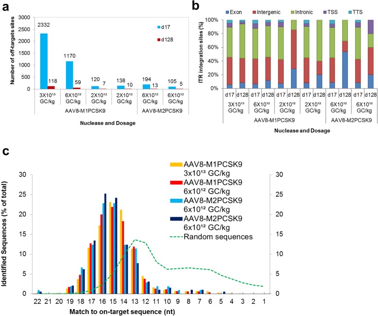
Fig. 2.
Analyzing on- and off-target activity of AAV8-M1PCSK9 and AAV8-M2PCSK9 in vivo. a. ITR-Seq-identified integration sites in liver samples treated with AAV8-M1PCSK9 and AAV8-M2PCSK9. Samples were collected on day 17 and 128 following vector administration. b. Functional annotation of ITR-identified integration sites. Here, we show the number of sites within exons, introns, intergenic regions, transcription start sites (TSS), and transcription termination sites (TTS). c. Distribution of ITR-integration sites on days 17/18 for two animals treated with either M1PCSK9 or M2PCSK9 (colored bars). Computationally generated random DNA sequences are represented by the green dotted line and are based on the number of nucleotides that match the intended target sequence (represented as a percent of all identified sites)

