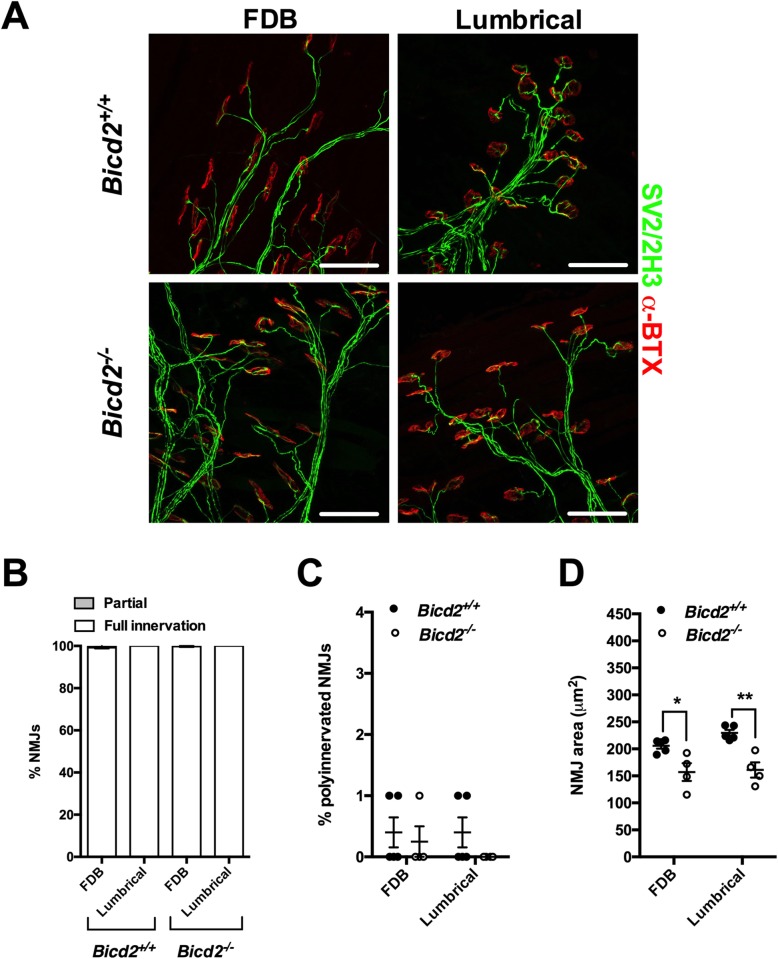
Fig. 3.
Normal NMJ analysis of Bicd2−/− mice at 21 days of age. a shows representative images of the NMJs of the FDB (flexor digitorum brevis) and feet lumbrical muscles stained with anti-SV2/2H3 antibodies (green) to visualise motor neurons and fluorescent alpha-bungarotoxin (red) to identify post-synaptic acetylcholine receptors on the muscle fibre surface. Scale bars = 50 μm. b shows the percentage of fully and partially innervated NMJs in Bicd2+/+ (n = 4) and Bicd2−/− (n = 4) mice. c shows the percentage of poly-innervated (measure of immaturity) NMJs between Bicd2+/+ (n = 4) and Bicd2−/− (n = 4) mice. d shows the area of the NMJ (area occupied by each single AchR cluster) in the FDB and lumbrical muscles in Bicd2+/+ (mean 205 and 230 μm2, respectively; n = 4) and Bicd2−/− (mean 157 and 161 μm2, respectively; n = 4) mice, (multiple t-tests corrected for multiple comparisons using the Holm-Sidak method, *p = 0.05, **p = 0.004). Error bars = SEM. The normal NMJ analysis suggests that there is no active denervation in Bicd2−/− mice at 21 days of age

