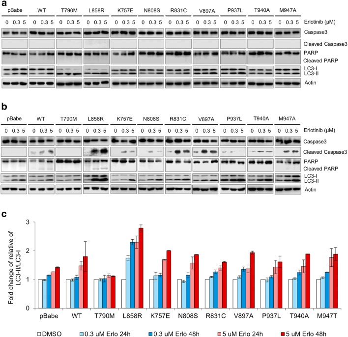
Fig. 7.
Erlotinib induces differential degree of induction apoptosis and autophagy among cells harboring different EGFRs. NIH-3T3 cells harboring EGFR WT and mutants were cultured in DMEM containing 4% FBS with different concentrations of erlotinib (0, 0.3, and 5 µM). Immunoblotting shows the effect of erlotinib on molecular markers of apoptosis and autophagy (caspase-3, PARP, LC3-I/II) after 24 h treatment (a) and 48 h treatment (b). Graphic presentations of the fold change of relative LC3-II/LC3-I in cells treated with erlotinib, presented as mean ± SEM (c)

