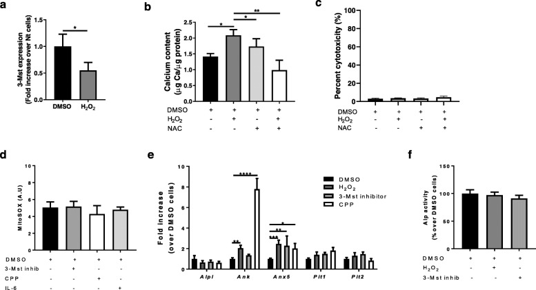
Fig. 4.
Oxidative stress regulates 3-MST expression, mineralization, and inflammation in chondrocytes. a qRT-PCR for 3-Mst gene expression in WT chondrocytes stimulated or not with 500 μM H2O2 for 4 h. n = 3. b Calcium content in chondrocytes monolayer incubated for 24 h with CPP and treated with vehicle (DMSO) or 500 μM H2O2 or with 1 mM NAC or with a combination of them. Calcium content is expressed in μg Calcium/μg protein. c LDH release in cell supernatant of chondrocytes from point (b). n = 3. d Mitochondrial ROS production (MitoSOX) in chondrocytes treated with vehicle (DMSO), or 50 μM 3-MST inhibitor or CPP or 10 ng/ml IL-6 for 1 h. n = 3. e qRT-PCR of the indicated genes in chondrocytes stimulated with vehicle (DMSO), or 500 μM H2O2, or 50 μM 3-MST inhibitor or CPP for 4 h. n = 3. f Alp activity in chondrocytes lysates treated with vehicle, or 500 μM H2O2, or 50 μM 3-MST inhibitor for 6 h. n = 3

