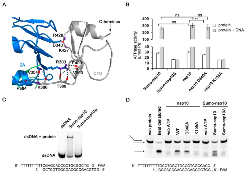
Figure 4.
Characterization of the PRRSV nsp10 CTD. (A) Close-up view of the domain interface between CTD and domain 2A. Residues engaged in interactions are shown as sticks. Domain colors are the same as in Figure 1A. (B) ATPase activities of PRRSV variants. The final concentrations of PRRSV nsp10 variants were 5 nM. (C) The binding affinity of PRRSV nsp10Δ to dsDNA is demonstrated through EMSA. (D) Helicase assay shows that PRRSV nsp10Δ cannot unwind dsDNA. No enzyme (w/o protein) and heat denatured controls are indicated. DNA strand with FAM label is marked with an asterisk. Error bars represent SD values from three separate experiments. ** P < 0.01; ns, not significant (Student’s unpaired t-test).

