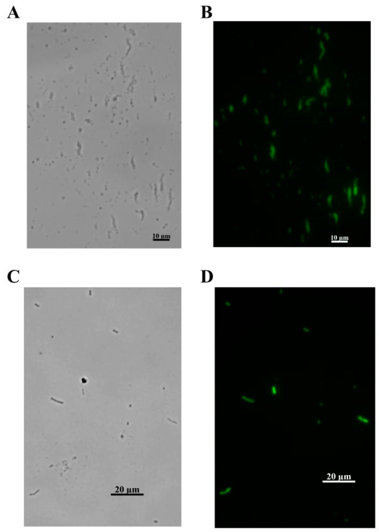
Figure 8.
FL micrographs of the stained A. baumannii. (A) The green FL channel of FITC-labelled gp52 stained bacterium. (B) The bright field of FITC-labelled gp52 stained bacterium. (C) The green FL channel of FITC-labelled gp53 stained bacterium. (D) The bright field of FITC-labelled gp53 stained bacterium.

