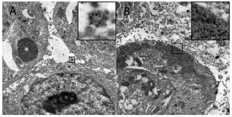
Figure 5.
Demonstration of the intracellular HIF-1α localization in persistently canine distemper virus infected DH82 cells as determined by immunoelectron microscopy. (A) HIF-1α was found within variably sized, round, moderately to highly electrondense vesicles (insert) and in large moderately electrondense vacuoles (*). Additionally, HIF-1α was detected often in the sub-membranous area of the cytoplasm (insert; B). Magnification 9000×.

