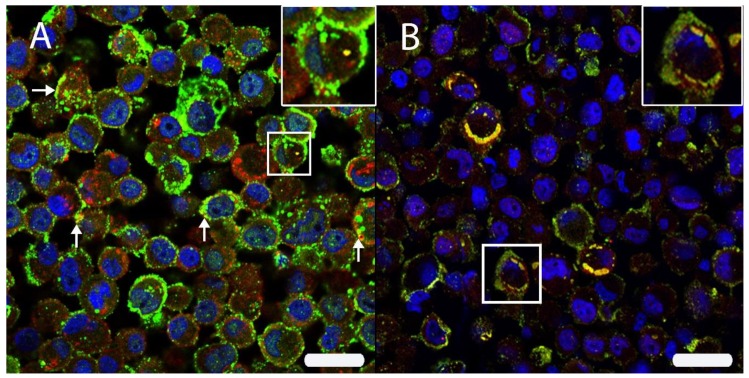
Figure 6.
(A) The intracellular HIF-1α localization was analyzed by double immunofluorescence with HIF-1α (Cy2, green) and CD63 (Cy3, red) in persistently canine distemper virus (CDV)-infected DH82 cells. Both proteins were localized within cell membranes and cytoplasm. Interestingly, an occasional co-expression (yellow) was noted (arrows; insert) using scanning confocal laser microscopy. (B) A double labeling directed against HIF-1α (Cy3, red) and the CDV nucleoprotein (CDV-NP; Cy2, green) revealed a frequent co-localization (yellow) beneath the cell membrane and within the perinuclear area (insert) of persistently CDV-infected DH82 cells. Nuclei were stained with bisbenzimide (blue). Bar = 20 µm.

