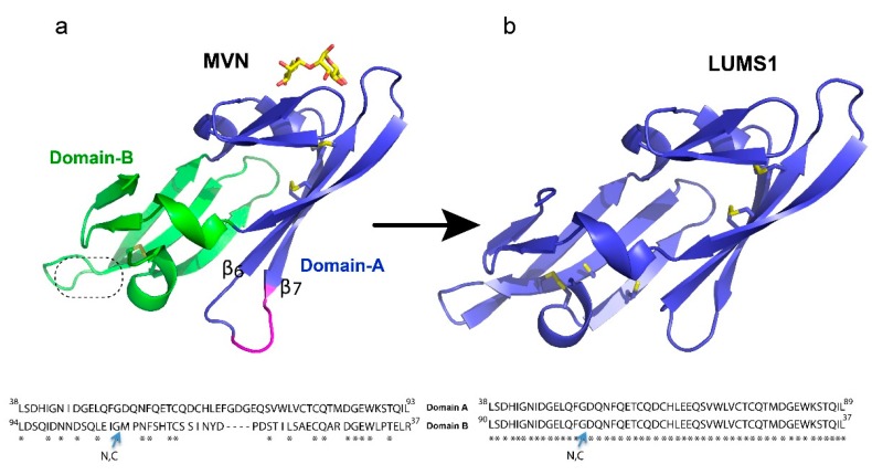
Figure 1.
Description of the protein design: (a) microvirin (MVN) structure (PDB ID 2YHH) shown in cartoon presentation with two structural domains colored blue and green while bound glycan is colored yellow. Insertion of four amino acids in domain-A as compared to domain-B is indicated in magenta. The second putative carbohydrate binding site is indicated by a dotted circle. (b) The homology-modeled structure of LUMS1 was created through SWISS-MODEL online tools using MVN as a template. Qualitative model energy analysis (QMEAN) scoring function was used to access the quality of the model. Side chains of all cysteine residues in both proteins are shown in gold sticks. Alignment of amino acid sequence of two domains of MVN and LUMS1 is shown at the bottom of the respective protein structure. N, C indicates N- and C-termini of the protein sequences.

