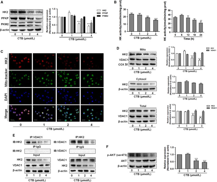
Figure 2.

CTB inhibited the expression of HK2 and promoted dissociation of hexokinase 2 from the mitochondria. SMMC‐7721 cells were treated with indicated concentrations of CTB (0, 1, 2 and 4 μmol/L). (A) Protein levels of HK2, PFKP and PKM2 were measured by Western blot analysis. (B) HK activity was detected via Hexokinase Activity Detection Kit at different time (0, 6, 12, 18, 24 h). (C) Representative fluorescence microscope images of SMMC‐7721 cells labelled with DAPI, HK2 antibody and Mito‐Tracker Green. Scale bar: 50 μm. (D) Mitochondrial and cytosolic fractions were isolated and subjected to Western blot analysis for the expression of HK2. (E) Immunoprecipitation assay showed the interaction of HK2 and VDAC1. (F) Protein levels of p‐AKT and AKT were measured by Western blot analysis. Data were presented as mean ± SD (n = 3); significance: *P < .05, **P < .01 and ***P < .001 vs control
